Dmitry A. Semchonok , Fotis L. Kyrilis , Farzad Hamdi , Panagiotis L. Kastritis
{"title":"Cryo-EM of a heterogeneous biochemical fraction elucidates multiple protein complexes from a multicellular thermophilic eukaryote","authors":"Dmitry A. Semchonok , Fotis L. Kyrilis , Farzad Hamdi , Panagiotis L. Kastritis","doi":"10.1016/j.yjsbx.2023.100094","DOIUrl":null,"url":null,"abstract":"<div><p>Biomolecular complexes and their interactions govern cellular structure and function. Understanding their architecture is a prerequisite for dissecting the cell's inner workings, but their higher-order assembly is often transient and challenging for structural analysis. Here, we performed cryo-EM on a single, highly heterogeneous biochemical fraction derived from <em>Chaetomium thermophilum</em> cell extracts to visualize the biomolecular content of the multicellular eukaryote. After cryo-EM single-particle image processing, results showed that a simultaneous three-dimensional structural characterization of multiple chemically diverse biomacromolecules is feasible. Namely, the thermophilic, eukaryotic complexes of (a) ATP citrate-lyase, (b) Hsp90, (c) 20S proteasome, (d) Hsp60 and (e) UDP-glucose pyrophosphorylase were characterized. In total, all five complexes have been structurally dissected in a thermophilic eukaryote in a total imaged sample area of 190.64 μm<sup>2</sup>, and two, in particular, 20S proteasome and Hsp60, exhibit side-chain resolution features. The <em>C. thermophilum</em> Hsp60 near-atomic model was resolved at 3.46 Å (FSC = 0.143) and shows a hinge-like conformational change of its equatorial domain, highly similar to the one previously shown for its bacterial orthologue, GroEL. This work demonstrates that cryo-EM of cell extracts will greatly accelerate the structural analysis of cellular complexes and provide unprecedented opportunities to annotate architectures of biomolecules in a holistic approach.</p></div>","PeriodicalId":17238,"journal":{"name":"Journal of Structural Biology: X","volume":"8 ","pages":"Article 100094"},"PeriodicalIF":5.1000,"publicationDate":"2023-08-09","publicationTypes":"Journal Article","fieldsOfStudy":null,"isOpenAccess":false,"openAccessPdf":"https://www.ncbi.nlm.nih.gov/pmc/articles/PMC10451023/pdf/","citationCount":"0","resultStr":null,"platform":"Semanticscholar","paperid":null,"PeriodicalName":"Journal of Structural Biology: X","FirstCategoryId":"1085","ListUrlMain":"https://www.sciencedirect.com/science/article/pii/S2590152423000107","RegionNum":0,"RegionCategory":null,"ArticlePicture":[],"TitleCN":null,"AbstractTextCN":null,"PMCID":null,"EPubDate":"","PubModel":"","JCR":"Q2","JCRName":"BIOCHEMISTRY & MOLECULAR BIOLOGY","Score":null,"Total":0}
引用次数: 0
Abstract
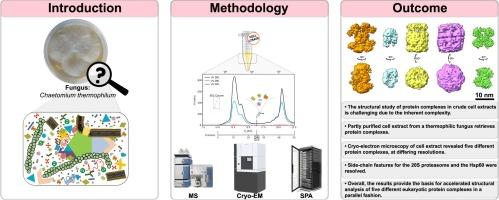
Biomolecular complexes and their interactions govern cellular structure and function. Understanding their architecture is a prerequisite for dissecting the cell's inner workings, but their higher-order assembly is often transient and challenging for structural analysis. Here, we performed cryo-EM on a single, highly heterogeneous biochemical fraction derived from Chaetomium thermophilum cell extracts to visualize the biomolecular content of the multicellular eukaryote. After cryo-EM single-particle image processing, results showed that a simultaneous three-dimensional structural characterization of multiple chemically diverse biomacromolecules is feasible. Namely, the thermophilic, eukaryotic complexes of (a) ATP citrate-lyase, (b) Hsp90, (c) 20S proteasome, (d) Hsp60 and (e) UDP-glucose pyrophosphorylase were characterized. In total, all five complexes have been structurally dissected in a thermophilic eukaryote in a total imaged sample area of 190.64 μm2, and two, in particular, 20S proteasome and Hsp60, exhibit side-chain resolution features. The C. thermophilum Hsp60 near-atomic model was resolved at 3.46 Å (FSC = 0.143) and shows a hinge-like conformational change of its equatorial domain, highly similar to the one previously shown for its bacterial orthologue, GroEL. This work demonstrates that cryo-EM of cell extracts will greatly accelerate the structural analysis of cellular complexes and provide unprecedented opportunities to annotate architectures of biomolecules in a holistic approach.
 求助内容:
求助内容: 应助结果提醒方式:


