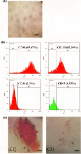
{"title":"Effects of MRI on stemness properties of Wharton's jelly-derived mesenchymal stem cells.","authors":"Mahnaz Tashakori, Fatemeh Asadi, Faezeh-Sadat Khorram, Azita Manshoori, Ali Hosseini-Chegeni, Fatemeh Mohseni Moghadam, Mahdieh Ahmadi Kamalabadi, Aliakbar Yousefi-Ahmadipour","doi":"10.1007/s10561-022-10052-2","DOIUrl":null,"url":null,"abstract":"<p><p>Mesenchymal stem cells (MSCs), derived from various tissues, are served as a promising source of cells in clinic and regenerative medicine. Umbilical cord-Wharton's jelly (WJ-MSCs)-derived MSCs exhibit advantages over those from adult tissues, such as no ethical concerns, shorter population doubling time, broad differentiation potential, readily available non-invasive source, prolonged maintenance of stemness properties. The aim of this study was to evaluate the effect of MRI (1.5 T, 10 min) on stemness gene expression patterns (OCT-4, SOX-2, NANOG) of WJ-MSCs. Additionally, we assessed cell viability, growth kinetics and apoptosis of WJ-MSCs after MRI treatment. The quantitative real-time reverse transcription polymerase chain reaction (qRT-PCR) data showed that transcript levels of SOX-2, NANOG in MRI-treated WJ-MSCs were increased 32- and 213-fold, respectively. MTT assay was performed at 24, 48, and 72 h post-treatment and the viability was not significantly different between the two groups. The doubling time of the MRI group was markedly higher than the control group. In addition, the colony formation ability of WJ-MSCs after MRI treatment significantly increased. Furthermore, no change in apoptosis was seen before or after MRI treatment. Our results suggest that the use of MRI can improve the quality of MSCs and enhance the efficacy of mesenchymal stem cell-based therapies.</p>","PeriodicalId":9723,"journal":{"name":"Cell and Tissue Banking","volume":"24 3","pages":"523-533"},"PeriodicalIF":1.4000,"publicationDate":"2023-09-01","publicationTypes":"Journal Article","fieldsOfStudy":null,"isOpenAccess":false,"openAccessPdf":"","citationCount":"0","resultStr":null,"platform":"Semanticscholar","paperid":null,"PeriodicalName":"Cell and Tissue Banking","FirstCategoryId":"5","ListUrlMain":"https://doi.org/10.1007/s10561-022-10052-2","RegionNum":4,"RegionCategory":"医学","ArticlePicture":[],"TitleCN":null,"AbstractTextCN":null,"PMCID":null,"EPubDate":"","PubModel":"","JCR":"Q4","JCRName":"CELL BIOLOGY","Score":null,"Total":0}
引用次数: 0
Abstract
Mesenchymal stem cells (MSCs), derived from various tissues, are served as a promising source of cells in clinic and regenerative medicine. Umbilical cord-Wharton's jelly (WJ-MSCs)-derived MSCs exhibit advantages over those from adult tissues, such as no ethical concerns, shorter population doubling time, broad differentiation potential, readily available non-invasive source, prolonged maintenance of stemness properties. The aim of this study was to evaluate the effect of MRI (1.5 T, 10 min) on stemness gene expression patterns (OCT-4, SOX-2, NANOG) of WJ-MSCs. Additionally, we assessed cell viability, growth kinetics and apoptosis of WJ-MSCs after MRI treatment. The quantitative real-time reverse transcription polymerase chain reaction (qRT-PCR) data showed that transcript levels of SOX-2, NANOG in MRI-treated WJ-MSCs were increased 32- and 213-fold, respectively. MTT assay was performed at 24, 48, and 72 h post-treatment and the viability was not significantly different between the two groups. The doubling time of the MRI group was markedly higher than the control group. In addition, the colony formation ability of WJ-MSCs after MRI treatment significantly increased. Furthermore, no change in apoptosis was seen before or after MRI treatment. Our results suggest that the use of MRI can improve the quality of MSCs and enhance the efficacy of mesenchymal stem cell-based therapies.
期刊介绍:
Cell and Tissue Banking provides a forum for disseminating information to scientists and clinicians involved in the banking and transplantation of cells and tissues. Cell and Tissue Banking is an international, peer-reviewed journal that publishes original papers in the following areas:
basic research concerning general aspects of tissue banking such as quality assurance and control of banked cells/tissues, effects of preservation and sterilisation methods on cells/tissues, biotechnology, etc.; clinical applications of banked cells/tissues; standards of practice in procurement, processing, storage and distribution of cells/tissues; ethical issues; medico-legal issues.
 求助内容:
求助内容: 应助结果提醒方式:


