Knockdown of histone deacetylase 9 attenuates sepsis-induced myocardial injury and inflammatory response.

IF 2.2 4区 农林科学 Q1 VETERINARY SCIENCES
Long Yang, Chunxue Wu, Ying Cui, Shimin Dong
{"title":"Knockdown of histone deacetylase 9 attenuates sepsis-induced myocardial injury and inflammatory response.","authors":"Long Yang, Chunxue Wu, Ying Cui, Shimin Dong","doi":"10.1538/expanim.22-0072","DOIUrl":null,"url":null,"abstract":"Myocardial cell damage is associated with apoptosis and excessive inflammatory response in sepsis. Histone deacetylases (HDACs) are implicated in the progression of heart diseases. This study aims to explore the role of histone deacetylase 9 (HDAC9) in sepsis-induced myocardial injury. Lipopolysaccharide (LPS)-induced Sprague Dawley rats and cardiomyocyte line H9C2 were used as models in vivo and in vitro. The results showed that HDAC9 was significantly upregulated after LPS stimulation, and HDAC9 knockdown remarkably improved cardiac function, as evidenced by decreased left ventricular internal diameter end diastole (LVEDD) and left ventricular internal diameter end systole (LVESD), and increased fractional shortening (FS)% and ejection fraction (EF)%. In addition, HDAC9 silencing alleviated release of inflammatory cytokines (tumor necrosis factor-α (TNF-α), IL-6 and IL-1β) and cardiomyocyte apoptosis in vivo and in vitro. Furthermore, HDAC9 inhibition was proved to suppress nuclear factor-kappa B (NF-κB) activation with reducing the levels of p-IκBα and p-p65, and p65 nuclear translocation. Additionally, interaction between miR-214-3p and HDAC9 was determined through bioinformatics analysis, RT-qPCR, western blot and dual luciferase reporter assay. Our data revealed that miR-214-3p directly targeted the 3’UTR of HDAC9. Our findings demonstrate that HDAC9 suppression ameliorates LPS-induced cardiac dysfunction by inhibiting the NF-κB signaling pathway and presents a promising therapeutic agent for the treatment of LPS-stimulated myocardial injury.","PeriodicalId":12102,"journal":{"name":"Experimental Animals","volume":"72 3","pages":"356-366"},"PeriodicalIF":2.2000,"publicationDate":"2023-08-07","publicationTypes":"Journal Article","fieldsOfStudy":null,"isOpenAccess":false,"openAccessPdf":"https://ftp.ncbi.nlm.nih.gov/pub/pmc/oa_pdf/4a/d6/expanim-72-356.PMC10435362.pdf","citationCount":"0","resultStr":null,"platform":"Semanticscholar","paperid":null,"PeriodicalName":"Experimental Animals","FirstCategoryId":"3","ListUrlMain":"https://doi.org/10.1538/expanim.22-0072","RegionNum":4,"RegionCategory":"农林科学","ArticlePicture":[],"TitleCN":null,"AbstractTextCN":null,"PMCID":null,"EPubDate":"","PubModel":"","JCR":"Q1","JCRName":"VETERINARY SCIENCES","Score":null,"Total":0}
引用次数: 0

Abstract

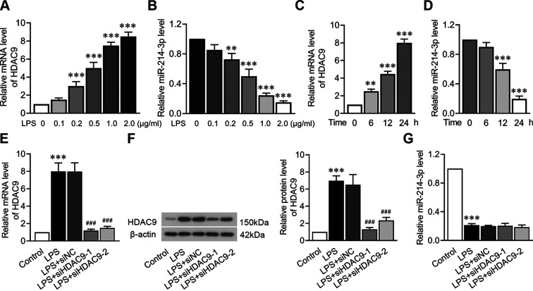
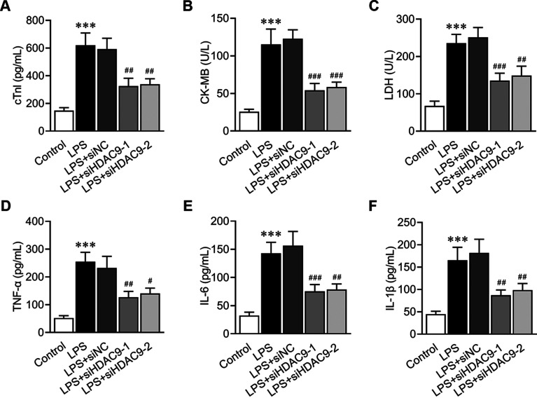
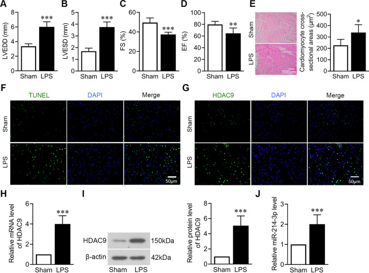
Myocardial cell damage is associated with apoptosis and excessive inflammatory response in sepsis. Histone deacetylases (HDACs) are implicated in the progression of heart diseases. This study aims to explore the role of histone deacetylase 9 (HDAC9) in sepsis-induced myocardial injury. Lipopolysaccharide (LPS)-induced Sprague Dawley rats and cardiomyocyte line H9C2 were used as models in vivo and in vitro. The results showed that HDAC9 was significantly upregulated after LPS stimulation, and HDAC9 knockdown remarkably improved cardiac function, as evidenced by decreased left ventricular internal diameter end diastole (LVEDD) and left ventricular internal diameter end systole (LVESD), and increased fractional shortening (FS)% and ejection fraction (EF)%. In addition, HDAC9 silencing alleviated release of inflammatory cytokines (tumor necrosis factor-α (TNF-α), IL-6 and IL-1β) and cardiomyocyte apoptosis in vivo and in vitro. Furthermore, HDAC9 inhibition was proved to suppress nuclear factor-kappa B (NF-κB) activation with reducing the levels of p-IκBα and p-p65, and p65 nuclear translocation. Additionally, interaction between miR-214-3p and HDAC9 was determined through bioinformatics analysis, RT-qPCR, western blot and dual luciferase reporter assay. Our data revealed that miR-214-3p directly targeted the 3’UTR of HDAC9. Our findings demonstrate that HDAC9 suppression ameliorates LPS-induced cardiac dysfunction by inhibiting the NF-κB signaling pathway and presents a promising therapeutic agent for the treatment of LPS-stimulated myocardial injury.
Abstract Image

Abstract Image

Abstract Image

组蛋白去乙酰化酶9的下调可减轻败血症引起的心肌损伤和炎症反应。
脓毒症患者心肌细胞损伤与细胞凋亡和过度炎症反应有关。组蛋白去乙酰化酶(hdac)与心脏病的进展有关。本研究旨在探讨组蛋白去乙酰化酶9 (HDAC9)在脓毒症致心肌损伤中的作用。以脂多糖(LPS)诱导的Sprague Dawley大鼠和心肌细胞系H9C2为体内和体外模型。结果显示,LPS刺激后HDAC9显著上调,HDAC9敲低可显著改善心功能,表现为左室舒张末期内径(LVEDD)和左室收缩末期内径(LVESD)降低,缩短分数(FS)%和射血分数(EF)%升高。此外,在体内和体外,HDAC9沉默可减轻炎症因子(肿瘤坏死因子-α (TNF-α)、IL-6和IL-1β)的释放和心肌细胞凋亡。此外,HDAC9抑制可以抑制核因子κB (NF-κB)的激活,降低p -κB α和p-p65的水平,以及p65核易位。此外,通过生物信息学分析、RT-qPCR、western blot和双荧光素酶报告基因检测来确定miR-214-3p与HDAC9的相互作用。我们的数据显示miR-214-3p直接靶向HDAC9的3'UTR。我们的研究结果表明,抑制HDAC9可通过抑制NF-κB信号通路改善脂多糖诱导的心功能障碍,为治疗脂多糖刺激的心肌损伤提供了一种有希望的治疗药物。
本文章由计算机程序翻译,如有差异,请以英文原文为准。
求助全文
约1分钟内获得全文 求助全文
来源期刊
Experimental Animals
Experimental Animals 生物-动物学
CiteScore
2.80
自引率
4.20%
发文量
2
审稿时长
3 months
期刊介绍: The aim of this international journal is to accelerate progress in laboratory animal experimentation and disseminate relevant information in related areas through publication of peer reviewed Original papers and Review articles. The journal covers basic to applied biomedical research centering around use of experimental animals and also covers topics related to experimental animals such as technology, management, and animal welfare.
×
引用
GB/T 7714-2015
复制
MLA
复制
APA
复制
导出至
BibTeX EndNote RefMan NoteFirst NoteExpress
×
提示
您的信息不完整,为了账户安全,请先补充。
现在去补充
×
提示
您因"违规操作"
具体请查看互助需知
我知道了
×
提示
确定
请完成安全验证×
copy
已复制链接
快去分享给好友吧!
我知道了
右上角分享
点击右上角分享
0
联系我们:info@booksci.cn Book学术提供免费学术资源搜索服务,方便国内外学者检索中英文文献。致力于提供最便捷和优质的服务体验。 Copyright © 2023 布克学术 All rights reserved.
京ICP备2023020795号-1
ghs 京公网安备 11010802042870号
Book学术文献互助
Book学术文献互助群
群 号:481959085
Book学术官方微信