Kelly Smart, Carme Uribe, Kimberly L Desmond, Sarah L Martin, Neil Vasdev, Antonio P Strafella
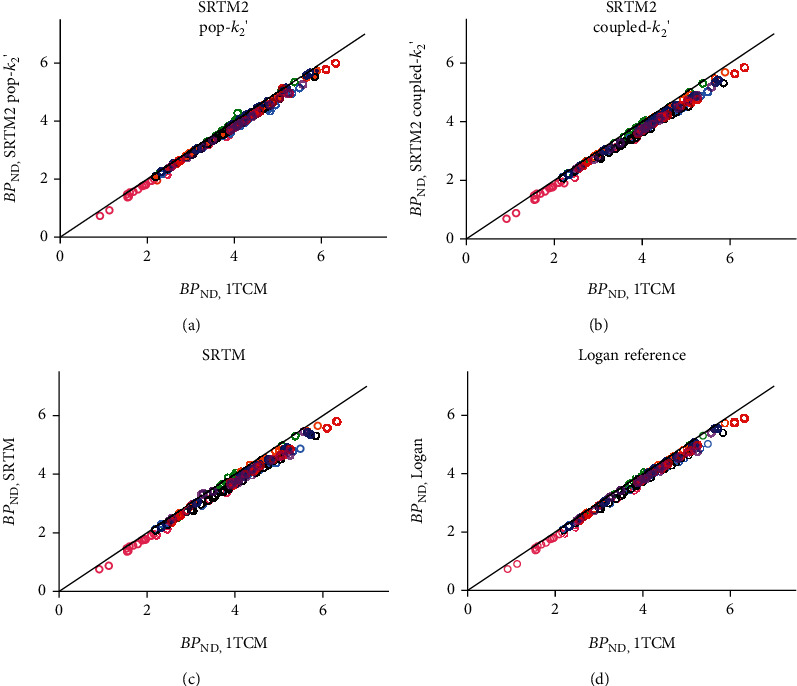
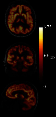
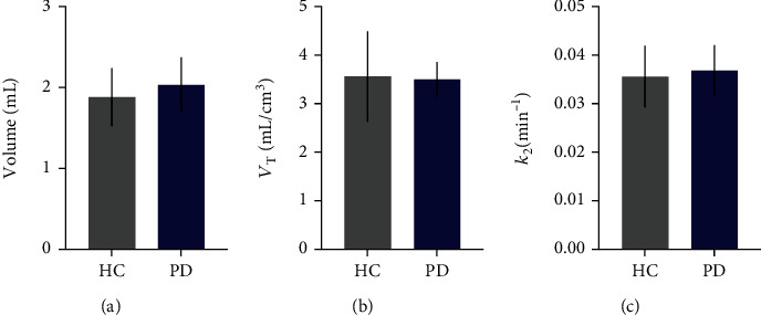
{"title":"Preliminary Assessment of Reference Region Quantification and Reduced Scanning Times for [<sup>18</sup>F]SynVesT-1 PET in Parkinson's Disease.","authors":"Kelly Smart, Carme Uribe, Kimberly L Desmond, Sarah L Martin, Neil Vasdev, Antonio P Strafella","doi":"10.1155/2023/1855985","DOIUrl":null,"url":null,"abstract":"<p><p>Synaptic density in the central nervous system can be measured <i>in vivo</i> using PET with [<sup>18</sup>F]SynVesT-1. While [<sup>18</sup>F]SynVesT-1 has been proven to be a powerful radiopharmaceutical for PET imaging of neurodegenerative disorders such as Parkinson's disease (PD), its currently validated acquisition and quantification protocols are invasive and technically challenging in these populations due to the arterial sampling and relatively long scanning times. The objectives of this work were to evaluate a noninvasive (reference tissue) quantification method for [<sup>18</sup>F]SynVesT-1 in PD patients and to determine the minimum scan time necessary for accurate quantification. [<sup>18</sup>F]SynVesT-1 PET scans were acquired in 5 patients with PD and 3 healthy control subjects for 120 min with arterial blood sampling. Quantification was performed using the one-tissue compartment model (1TCM) with arterial input function, as well as with the simplified reference tissue model (SRTM) to estimate binding potential (BP<sub>ND</sub>) using centrum semiovale (CS) as a reference region. The SRTM2 method was used with <i>k</i><sub>2</sub>' fixed to either a sample average value (0.037 min<sup>-1</sup>) or a value estimated first through coupled fitting across regions for each participant. Direct SRTM estimation and the Logan reference region graphical method were also evaluated. There were no significant group differences in CS volume, radiotracer uptake, or efflux (<i>ps</i> > 0.47). Each fitting method produced BP<sub>ND</sub> estimates in close agreement with those derived from the 1TCM (subject <i>R</i><sup>2</sup>s > 0.98, bias < 10%), with no difference in bias between the control and PD groups. With SRTM2, BP<sub>ND</sub> estimates from truncated scan data as short as 80 min produced values in excellent agreement with the data from the full 120 min scans (bias < 6%). While these are preliminary results from a small sample of patients with PD (<i>n</i> = 5), this work suggests that accurate synaptic density quantification may be performed without blood sampling and with scan time under 90 minutes. If further validated, these simplified procedures for [<sup>18</sup>F]SynVesT-1 PET quantification can facilitate its application as a clinical research imaging technology and allow for larger study samples and include a broader scope of patients including those with neurodegenerative diseases.</p>","PeriodicalId":18855,"journal":{"name":"Molecular Imaging","volume":"2023 ","pages":"1855985"},"PeriodicalIF":2.4000,"publicationDate":"2023-08-11","publicationTypes":"Journal Article","fieldsOfStudy":null,"isOpenAccess":false,"openAccessPdf":"https://www.ncbi.nlm.nih.gov/pmc/articles/PMC10445483/pdf/","citationCount":"0","resultStr":null,"platform":"Semanticscholar","paperid":null,"PeriodicalName":"Molecular Imaging","FirstCategoryId":"3","ListUrlMain":"https://doi.org/10.1155/2023/1855985","RegionNum":4,"RegionCategory":"医学","ArticlePicture":[],"TitleCN":null,"AbstractTextCN":null,"PMCID":null,"EPubDate":"2023/1/1 0:00:00","PubModel":"eCollection","JCR":"Q3","JCRName":"BIOCHEMICAL RESEARCH METHODS","Score":null,"Total":0}
引用次数: 0
Abstract
Synaptic density in the central nervous system can be measured in vivo using PET with [18F]SynVesT-1. While [18F]SynVesT-1 has been proven to be a powerful radiopharmaceutical for PET imaging of neurodegenerative disorders such as Parkinson's disease (PD), its currently validated acquisition and quantification protocols are invasive and technically challenging in these populations due to the arterial sampling and relatively long scanning times. The objectives of this work were to evaluate a noninvasive (reference tissue) quantification method for [18F]SynVesT-1 in PD patients and to determine the minimum scan time necessary for accurate quantification. [18F]SynVesT-1 PET scans were acquired in 5 patients with PD and 3 healthy control subjects for 120 min with arterial blood sampling. Quantification was performed using the one-tissue compartment model (1TCM) with arterial input function, as well as with the simplified reference tissue model (SRTM) to estimate binding potential (BPND) using centrum semiovale (CS) as a reference region. The SRTM2 method was used with k2' fixed to either a sample average value (0.037 min-1) or a value estimated first through coupled fitting across regions for each participant. Direct SRTM estimation and the Logan reference region graphical method were also evaluated. There were no significant group differences in CS volume, radiotracer uptake, or efflux (ps > 0.47). Each fitting method produced BPND estimates in close agreement with those derived from the 1TCM (subject R2s > 0.98, bias < 10%), with no difference in bias between the control and PD groups. With SRTM2, BPND estimates from truncated scan data as short as 80 min produced values in excellent agreement with the data from the full 120 min scans (bias < 6%). While these are preliminary results from a small sample of patients with PD (n = 5), this work suggests that accurate synaptic density quantification may be performed without blood sampling and with scan time under 90 minutes. If further validated, these simplified procedures for [18F]SynVesT-1 PET quantification can facilitate its application as a clinical research imaging technology and allow for larger study samples and include a broader scope of patients including those with neurodegenerative diseases.
Molecular ImagingBiochemistry, Genetics and Molecular Biology-Biotechnology
自引率
3.60%
发文量
21
期刊介绍:
Molecular Imaging is a peer-reviewed, open access journal highlighting the breadth of molecular imaging research from basic science to preclinical studies to human applications. This serves both the scientific and clinical communities by disseminating novel results and concepts relevant to the biological study of normal and disease processes in both basic and translational studies ranging from mice to humans.
 求助内容:
求助内容: 应助结果提醒方式:


