Rachael E Redgrave, Emily Dookun, Laura K Booth, Maria Camacho Encina, Omowumi Folaranmi, Simon Tual-Chalot, Jason H Gill, W Andrew Owens, Ioakim Spyridopoulos, João F Passos, Gavin D Richardson
{"title":"Senescent cardiomyocytes contribute to cardiac dysfunction following myocardial infarction.","authors":"Rachael E Redgrave, Emily Dookun, Laura K Booth, Maria Camacho Encina, Omowumi Folaranmi, Simon Tual-Chalot, Jason H Gill, W Andrew Owens, Ioakim Spyridopoulos, João F Passos, Gavin D Richardson","doi":"10.1038/s41514-023-00113-5","DOIUrl":null,"url":null,"abstract":"<p><p>Myocardial infarction is a leading cause of morbidity and mortality. While reperfusion is now standard therapy, pathological remodelling leading to heart failure remains a clinical problem. Cellular senescence has been shown to contribute to disease pathophysiology and treatment with the senolytic navitoclax attenuates inflammation, reduces adverse myocardial remodelling and results in improved functional recovery. However, it remains unclear which senescent cell populations contribute to these processes. To identify whether senescent cardiomyocytes contribute to disease pathophysiology post-myocardial infarction, we established a transgenic model in which p16 (CDKN2A) expression was specifically knocked-out in the cardiomyocyte population. Following myocardial infarction, mice lacking cardiomyocyte p16 expression demonstrated no difference in cardiomyocyte hypertrophy but exhibited improved cardiac function and significantly reduced scar size in comparison to control animals. This data demonstrates that senescent cardiomyocytes participate in pathological myocardial remodelling. Importantly, inhibition of cardiomyocyte senescence led to reduced senescence-associated inflammation and decreased senescence-associated markers within other myocardial lineages, consistent with the hypothesis that cardiomyocytes promote pathological remodelling by spreading senescence to other cell-types. Collectively this study presents the demonstration that senescent cardiomyocytes are major contributors to myocardial remodelling and dysfunction following a myocardial infarction. Therefore, to maximise the potential for clinical translation, it is important to further understand the mechanisms underlying cardiomyocyte senescence and how to optimise senolytic strategies to target this cell lineage.</p>","PeriodicalId":19348,"journal":{"name":"npj Aging","volume":"9 1","pages":"15"},"PeriodicalIF":0.0000,"publicationDate":"2023-06-14","publicationTypes":"Journal Article","fieldsOfStudy":null,"isOpenAccess":false,"openAccessPdf":"https://www.ncbi.nlm.nih.gov/pmc/articles/PMC10267185/pdf/","citationCount":"0","resultStr":null,"platform":"Semanticscholar","paperid":null,"PeriodicalName":"npj Aging","FirstCategoryId":"1085","ListUrlMain":"https://doi.org/10.1038/s41514-023-00113-5","RegionNum":0,"RegionCategory":null,"ArticlePicture":[],"TitleCN":null,"AbstractTextCN":null,"PMCID":null,"EPubDate":"","PubModel":"","JCR":"","JCRName":"","Score":null,"Total":0}
引用次数: 0
Abstract
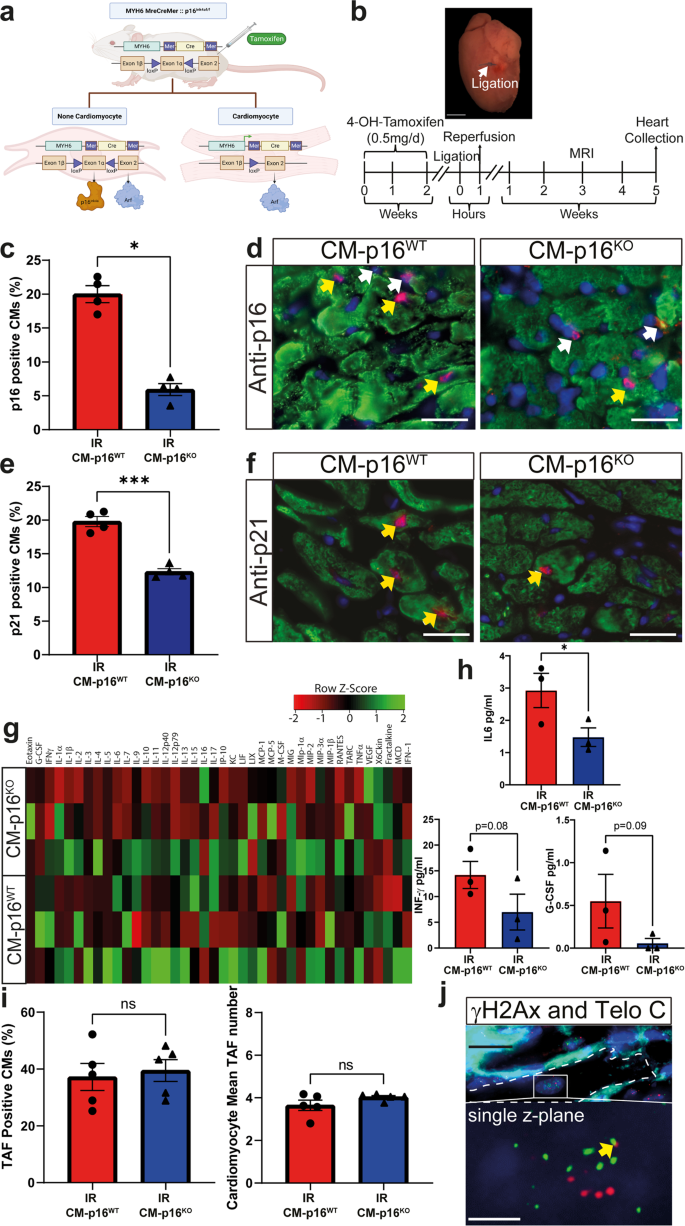
Myocardial infarction is a leading cause of morbidity and mortality. While reperfusion is now standard therapy, pathological remodelling leading to heart failure remains a clinical problem. Cellular senescence has been shown to contribute to disease pathophysiology and treatment with the senolytic navitoclax attenuates inflammation, reduces adverse myocardial remodelling and results in improved functional recovery. However, it remains unclear which senescent cell populations contribute to these processes. To identify whether senescent cardiomyocytes contribute to disease pathophysiology post-myocardial infarction, we established a transgenic model in which p16 (CDKN2A) expression was specifically knocked-out in the cardiomyocyte population. Following myocardial infarction, mice lacking cardiomyocyte p16 expression demonstrated no difference in cardiomyocyte hypertrophy but exhibited improved cardiac function and significantly reduced scar size in comparison to control animals. This data demonstrates that senescent cardiomyocytes participate in pathological myocardial remodelling. Importantly, inhibition of cardiomyocyte senescence led to reduced senescence-associated inflammation and decreased senescence-associated markers within other myocardial lineages, consistent with the hypothesis that cardiomyocytes promote pathological remodelling by spreading senescence to other cell-types. Collectively this study presents the demonstration that senescent cardiomyocytes are major contributors to myocardial remodelling and dysfunction following a myocardial infarction. Therefore, to maximise the potential for clinical translation, it is important to further understand the mechanisms underlying cardiomyocyte senescence and how to optimise senolytic strategies to target this cell lineage.

 求助内容:
求助内容: 应助结果提醒方式:


