Ga-Eun Kim, Su-Jee Park, Yeong Jin Kim, Seul-Kee Kim, Tae-Young Jung
{"title":"Radiologic Follow-up of Ruptured Arachnoid Cysts With or Without Hemorrhage: Five Case Reports and a Review of the Literature.","authors":"Ga-Eun Kim, Su-Jee Park, Yeong Jin Kim, Seul-Kee Kim, Tae-Young Jung","doi":"10.14791/btrt.2023.0013","DOIUrl":null,"url":null,"abstract":"<p><p>Arachnoid cysts are usually asymptomatic and discovered incidentally. However, cysts may occasionally rupture because of minor head trauma. We describe the radiologic follow-up of 5 patients with ruptured arachnoid cysts featuring spontaneous resolution, subdural hygroma formation, and cystic and subdural hemorrhage. From January 2004 through July 2020, 5 patients (1.3%) with ruptured arachnoid cysts were evaluated out of 388 patients with arachnoid cysts encountered at our institution at that time. The 5 patients were all male, and they ranged in age from 6-17 years (median, 12 years). The median duration of radiologic follow-up was 3.5 years (range, 2.3-10.1 years). All of the ruptured arachnoid cysts were overlying the temporal lobe with Galassi type II. The median cyst diameter was 4.9 cm (range, 4.4-8.9 cm). Four patients had a history of recent minor head trauma. There were no particular neurologic symptoms in their past medical history in all patients. In the follow-up, two patients' cysts resolved spontaneously without hemorrhage. One patient's cyst resolved post-burr-hole drainage for chronic subdural hemorrhage. Another patient, whose cyst led to a hemorrhage and chronic subdural hemorrhage, recovered following a craniotomy, hematoma removal, and cyst fenestration. Another patient, presenting with hygroma, cystic hemorrhage, and chronic subdural hemorrhage, was treated with burr-hole drainage. Three patients recovered postoperatively. Arachnoid cysts rarely rupture, and surgical intervention is required for some cases associated with hemorrhage. Postoperatively, all patients had good outcomes without complications in this series.</p>","PeriodicalId":72453,"journal":{"name":"Brain tumor research and treatment","volume":"11 3","pages":"210-215"},"PeriodicalIF":0.0000,"publicationDate":"2023-07-01","publicationTypes":"Journal Article","fieldsOfStudy":null,"isOpenAccess":false,"openAccessPdf":"https://ftp.ncbi.nlm.nih.gov/pub/pmc/oa_pdf/8f/f4/btrt-11-210.PMC10409616.pdf","citationCount":"0","resultStr":null,"platform":"Semanticscholar","paperid":null,"PeriodicalName":"Brain tumor research and treatment","FirstCategoryId":"1085","ListUrlMain":"https://doi.org/10.14791/btrt.2023.0013","RegionNum":0,"RegionCategory":null,"ArticlePicture":[],"TitleCN":null,"AbstractTextCN":null,"PMCID":null,"EPubDate":"","PubModel":"","JCR":"","JCRName":"","Score":null,"Total":0}
引用次数: 0
Abstract
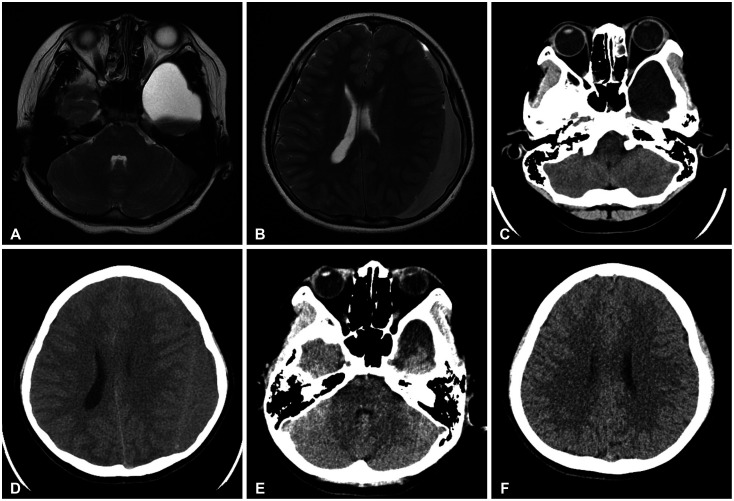
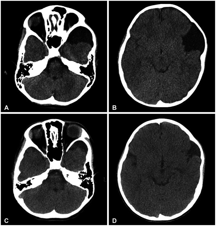
Arachnoid cysts are usually asymptomatic and discovered incidentally. However, cysts may occasionally rupture because of minor head trauma. We describe the radiologic follow-up of 5 patients with ruptured arachnoid cysts featuring spontaneous resolution, subdural hygroma formation, and cystic and subdural hemorrhage. From January 2004 through July 2020, 5 patients (1.3%) with ruptured arachnoid cysts were evaluated out of 388 patients with arachnoid cysts encountered at our institution at that time. The 5 patients were all male, and they ranged in age from 6-17 years (median, 12 years). The median duration of radiologic follow-up was 3.5 years (range, 2.3-10.1 years). All of the ruptured arachnoid cysts were overlying the temporal lobe with Galassi type II. The median cyst diameter was 4.9 cm (range, 4.4-8.9 cm). Four patients had a history of recent minor head trauma. There were no particular neurologic symptoms in their past medical history in all patients. In the follow-up, two patients' cysts resolved spontaneously without hemorrhage. One patient's cyst resolved post-burr-hole drainage for chronic subdural hemorrhage. Another patient, whose cyst led to a hemorrhage and chronic subdural hemorrhage, recovered following a craniotomy, hematoma removal, and cyst fenestration. Another patient, presenting with hygroma, cystic hemorrhage, and chronic subdural hemorrhage, was treated with burr-hole drainage. Three patients recovered postoperatively. Arachnoid cysts rarely rupture, and surgical intervention is required for some cases associated with hemorrhage. Postoperatively, all patients had good outcomes without complications in this series.


 求助内容:
求助内容: 应助结果提醒方式:


