Koji Itagaki, Kanae K Miyake, Minori Tanoue, Tae Oishi, Masako Kataoka, Masahiro Kawashima, Masakazu Toi, Yuji Nakamoto
{"title":"Feasibility of Dedicated Breast Positron Emission Tomography Image Denoising Using a Residual Neural Network.","authors":"Koji Itagaki, Kanae K Miyake, Minori Tanoue, Tae Oishi, Masako Kataoka, Masahiro Kawashima, Masakazu Toi, Yuji Nakamoto","doi":"10.22038/AOJNMB.2023.71598.1501","DOIUrl":null,"url":null,"abstract":"<p><strong>Objectives: </strong>This study aimed to create a deep learning (DL)-based denoising model using a residual neural network (Res-Net) trained to reduce noise in ring-type dedicated breast positron emission tomography (dbPET) images acquired in about half the emission time, and to evaluate the feasibility and the effectiveness of the model in terms of its noise reduction performance and preservation of quantitative values compared to conventional post-image filtering techniques.</p><p><strong>Methods: </strong>Low-count (LC) and full-count (FC) PET images with acquisition durations of 3 and 7 minutes, respectively, were reconstructed. A Res-Net was trained to create a noise reduction model using fifteen patients' data. The inputs to the network were LC images and its outputs were denoised PET (LC + DL) images, which should resemble FC images. To evaluate the LC + DL images, Gaussian and non-local mean (NLM) filters were applied to the LC images (LC + Gaussian and LC + NLM, respectively). To create reference images, a Gaussian filter was applied to the FC images (FC + Gaussian). The usefulness of our denoising model was objectively and visually evaluated using test data set of thirteen patients. The coefficient of variation (CV) of background fibroglandular tissue or fat tissue were measured to evaluate the performance of the noise reduction. The SUV<sub>max</sub> and SUV<sub>peak</sub> of lesions were also measured. The agreement of the SUV measurements was evaluated by Bland-Altman plots.</p><p><strong>Results: </strong>The CV of background fibroglandular tissue in the LC + DL images was significantly lower (9.10<math><mo>±</mo></math>2.76) than the CVs in the LC (13.60<math><mo>±</mo></math> 3.66) and LC + Gaussian images (11.51<math><mo>±</mo></math> 3.56). No significant difference was observed in both SUV<sub>max</sub> and SUV<sub>peak</sub> of lesions between LC + DL and reference images. For the visual assessment, the smoothness rating for the LC + DL images was significantly better than that for the other images except for the reference images.</p><p><strong>Conclusion: </strong>Our model reduced the noise in dbPET images acquired in about half the emission time while preserving quantitative values of lesions. This study demonstrates that machine learning is feasible and potentially performs better than conventional post-image filtering in dbPET denoising.</p>","PeriodicalId":8503,"journal":{"name":"Asia Oceania Journal of Nuclear Medicine and Biology","volume":null,"pages":null},"PeriodicalIF":0.0000,"publicationDate":"2023-01-01","publicationTypes":"Journal Article","fieldsOfStudy":null,"isOpenAccess":false,"openAccessPdf":"https://www.ncbi.nlm.nih.gov/pmc/articles/PMC10261694/pdf/","citationCount":"0","resultStr":null,"platform":"Semanticscholar","paperid":null,"PeriodicalName":"Asia Oceania Journal of Nuclear Medicine and Biology","FirstCategoryId":"1085","ListUrlMain":"https://doi.org/10.22038/AOJNMB.2023.71598.1501","RegionNum":0,"RegionCategory":null,"ArticlePicture":[],"TitleCN":null,"AbstractTextCN":null,"PMCID":null,"EPubDate":"","PubModel":"","JCR":"Q3","JCRName":"Medicine","Score":null,"Total":0}
引用次数: 0
Abstract
Objectives: This study aimed to create a deep learning (DL)-based denoising model using a residual neural network (Res-Net) trained to reduce noise in ring-type dedicated breast positron emission tomography (dbPET) images acquired in about half the emission time, and to evaluate the feasibility and the effectiveness of the model in terms of its noise reduction performance and preservation of quantitative values compared to conventional post-image filtering techniques.
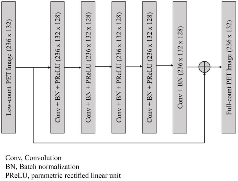
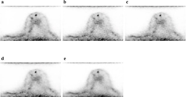
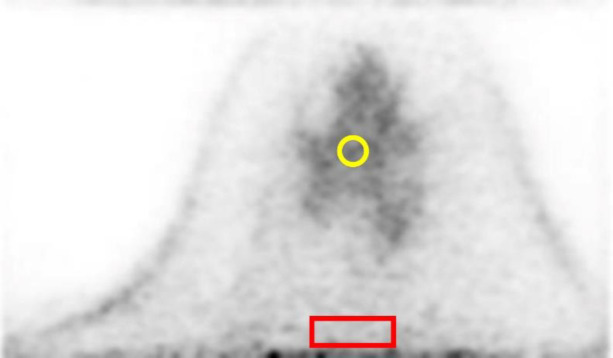
Methods: Low-count (LC) and full-count (FC) PET images with acquisition durations of 3 and 7 minutes, respectively, were reconstructed. A Res-Net was trained to create a noise reduction model using fifteen patients' data. The inputs to the network were LC images and its outputs were denoised PET (LC + DL) images, which should resemble FC images. To evaluate the LC + DL images, Gaussian and non-local mean (NLM) filters were applied to the LC images (LC + Gaussian and LC + NLM, respectively). To create reference images, a Gaussian filter was applied to the FC images (FC + Gaussian). The usefulness of our denoising model was objectively and visually evaluated using test data set of thirteen patients. The coefficient of variation (CV) of background fibroglandular tissue or fat tissue were measured to evaluate the performance of the noise reduction. The SUVmax and SUVpeak of lesions were also measured. The agreement of the SUV measurements was evaluated by Bland-Altman plots.
Results: The CV of background fibroglandular tissue in the LC + DL images was significantly lower (9.102.76) than the CVs in the LC (13.60 3.66) and LC + Gaussian images (11.51 3.56). No significant difference was observed in both SUVmax and SUVpeak of lesions between LC + DL and reference images. For the visual assessment, the smoothness rating for the LC + DL images was significantly better than that for the other images except for the reference images.
Conclusion: Our model reduced the noise in dbPET images acquired in about half the emission time while preserving quantitative values of lesions. This study demonstrates that machine learning is feasible and potentially performs better than conventional post-image filtering in dbPET denoising.

 求助内容:
求助内容: 应助结果提醒方式:


