Sergio M de Almeida, Miriam Perlingeiro Beltrame, Bin Tang, Indianara Rotta, Ian Abramson, Florin Vaida, Rachel Schrier, Ronald J Ellis
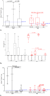
{"title":"Cerebrospinal fluid CD14<sup>++</sup>CD16<sup>+</sup> monocytes in HIV-1 subtype C compared with subtype B.","authors":"Sergio M de Almeida, Miriam Perlingeiro Beltrame, Bin Tang, Indianara Rotta, Ian Abramson, Florin Vaida, Rachel Schrier, Ronald J Ellis","doi":"10.1007/s13365-023-01137-z","DOIUrl":null,"url":null,"abstract":"<p><p>CD14<sup>++</sup>CD16<sup>+</sup> monocytes are susceptible to HIV-1 infection, and cross the blood-brain barrier. HIV-1 subtype C (HIV-1C) shows reduced Tat protein chemoattractant activity compared to HIV-1B, which might influence monocyte trafficking into the CNS. We hypothesized that the proportion of monocytes in CSF in HIV-1C is lower than HIV-1B group. We sought to assess differences in monocyte proportions in cerebrospinal fluid (CSF) and peripheral blood (PB) between people with HIV (PWH) and without HIV (PWoH), and by HIV-1B and -C subtypes. Immunophenotyping was performed by flow cytometry, monocytes were analyzed within CD45 + and CD64 + gated regions and classified in classical (CD14<sup>++</sup>CD16<sup>-</sup>), intermediate (CD14<sup>++</sup>CD16<sup>+</sup>), and non-classical (CD14<sup>low</sup>CD16<sup>+</sup>). Among PWH, the median [IQR] CD4 nadir was 219 [32-531] cell/mm<sup>3</sup>; plasma HIV RNA (log<sub>10</sub>) was 1.60 [1.60-3.21], and 68% were on antiretroviral therapy (ART). Participants with HIV-1C and -B were comparable in terms of age, duration of infection, CD4 nadir, plasma HIV RNA, and ART. The proportion of CSF CD14<sup>++</sup>CD16<sup>+</sup> monocytes was higher in participants with HIV-1C than those with HIV-1B [2.00(0.00-2.80) vs. 0.00(0.00-0.60) respectively, p = 0.03 after BH correction p = 0.10]. Despite viral suppression, the proportion of total monocytes in PB increased in PWH, due to the increase in CD14<sup>++</sup>CD16<sup>+</sup> and CD14<sup>low</sup>CD16<sup>+</sup> monocytes. The HIV-1C Tat substitution (C30S31) did not interfere with the migration of CD14<sup>++</sup>CD16<sup>+</sup> monocytes to the CNS. This is the first study to evaluate these monocytes in the CSF and PB and compare their proportions according to HIV subtype.</p>","PeriodicalId":16665,"journal":{"name":"Journal of NeuroVirology","volume":null,"pages":null},"PeriodicalIF":2.3000,"publicationDate":"2023-06-01","publicationTypes":"Journal Article","fieldsOfStudy":null,"isOpenAccess":false,"openAccessPdf":"https://www.ncbi.nlm.nih.gov/pmc/articles/PMC10769008/pdf/","citationCount":"0","resultStr":null,"platform":"Semanticscholar","paperid":null,"PeriodicalName":"Journal of NeuroVirology","FirstCategoryId":"3","ListUrlMain":"https://doi.org/10.1007/s13365-023-01137-z","RegionNum":4,"RegionCategory":"医学","ArticlePicture":[],"TitleCN":null,"AbstractTextCN":null,"PMCID":null,"EPubDate":"2023/5/23 0:00:00","PubModel":"Epub","JCR":"Q3","JCRName":"NEUROSCIENCES","Score":null,"Total":0}
引用次数: 0
Abstract
CD14++CD16+ monocytes are susceptible to HIV-1 infection, and cross the blood-brain barrier. HIV-1 subtype C (HIV-1C) shows reduced Tat protein chemoattractant activity compared to HIV-1B, which might influence monocyte trafficking into the CNS. We hypothesized that the proportion of monocytes in CSF in HIV-1C is lower than HIV-1B group. We sought to assess differences in monocyte proportions in cerebrospinal fluid (CSF) and peripheral blood (PB) between people with HIV (PWH) and without HIV (PWoH), and by HIV-1B and -C subtypes. Immunophenotyping was performed by flow cytometry, monocytes were analyzed within CD45 + and CD64 + gated regions and classified in classical (CD14++CD16-), intermediate (CD14++CD16+), and non-classical (CD14lowCD16+). Among PWH, the median [IQR] CD4 nadir was 219 [32-531] cell/mm3; plasma HIV RNA (log10) was 1.60 [1.60-3.21], and 68% were on antiretroviral therapy (ART). Participants with HIV-1C and -B were comparable in terms of age, duration of infection, CD4 nadir, plasma HIV RNA, and ART. The proportion of CSF CD14++CD16+ monocytes was higher in participants with HIV-1C than those with HIV-1B [2.00(0.00-2.80) vs. 0.00(0.00-0.60) respectively, p = 0.03 after BH correction p = 0.10]. Despite viral suppression, the proportion of total monocytes in PB increased in PWH, due to the increase in CD14++CD16+ and CD14lowCD16+ monocytes. The HIV-1C Tat substitution (C30S31) did not interfere with the migration of CD14++CD16+ monocytes to the CNS. This is the first study to evaluate these monocytes in the CSF and PB and compare their proportions according to HIV subtype.
期刊介绍:
The Journal of NeuroVirology (JNV) provides a unique platform for the publication of high-quality basic science and clinical studies on the molecular biology and pathogenesis of viral infections of the nervous system, and for reporting on the development of novel therapeutic strategies using neurotropic viral vectors. The Journal also emphasizes publication of non-viral infections that affect the central nervous system. The Journal publishes original research articles, reviews, case reports, coverage of various scientific meetings, along with supplements and special issues on selected subjects.
The Journal is currently accepting submissions of original work from the following basic and clinical research areas: Aging & Neurodegeneration, Apoptosis, CNS Signal Transduction, Emerging CNS Infections, Molecular Virology, Neural-Immune Interaction, Novel Diagnostics, Novel Therapeutics, Stem Cell Biology, Transmissable Encephalopathies/Prion, Vaccine Development, Viral Genomics, Viral Neurooncology, Viral Neurochemistry, Viral Neuroimmunology, Viral Neuropharmacology.
 求助内容:
求助内容: 应助结果提醒方式:


