Enrique Jimenez Hakim, Luis Garcia Rairan, Julian Guzman, Yessid Araque
{"title":"迷走神经球引起的双侧视觉障碍:一个例证性病例。","authors":"Enrique Jimenez Hakim, Luis Garcia Rairan, Julian Guzman, Yessid Araque","doi":"10.3171/CASE23145","DOIUrl":null,"url":null,"abstract":"<p><strong>Background: </strong>A glomus vagale tumor is an infrequent paraganglioma primarily characterized by auditory symptoms, cranial nerve involvement, or autonomic symptoms. However, visual involvement is not commonly observed, and to date, no cases have been reported in the literature.</p><p><strong>Observations: </strong>The case involves a 62-year-old female patient with a history of right carotid body tumor resection. She presented to the emergency department with a sudden decrease in visual acuity and bitemporal hemianopsia, accompanied by a left parietal headache. Initial brain magnetic resonance imaging (MRI) revealed a pituitary macroadenoma, which was completely resected. However, postoperatively, the patient developed left amaurosis. Subsequent brain MRI showed the presence of hemostatic material mixed with blood in the sellar region, causing displacement of the optic chiasm. A repeat intervention was performed, identifying bleeding from both cavernous sinuses. Head and neck angiography demonstrated a right glomus vagale tumor with abundant blood drainage into the right cavernous sinus. Embolization of the glomus vagale tumor was performed, resulting in no further bleeding and improvement of symptoms.</p><p><strong>Lessons: </strong>The aim of this case report is to describe a rare occurrence of bilateral visual disturbances caused by bleeding in both cavernous sinuses due to venous hypertension caused by a right glomus vagale tumor.</p>","PeriodicalId":16554,"journal":{"name":"Journal of Neurosurgery: Case Lessons","volume":"6 6","pages":""},"PeriodicalIF":0.0000,"publicationDate":"2023-08-07","publicationTypes":"Journal Article","fieldsOfStudy":null,"isOpenAccess":false,"openAccessPdf":"https://ftp.ncbi.nlm.nih.gov/pub/pmc/oa_pdf/f5/a5/CASE23145.PMC10555592.pdf","citationCount":"0","resultStr":"{\"title\":\"Bilateral visual disturbances caused by a glomus vagale: illustrative case.\",\"authors\":\"Enrique Jimenez Hakim, Luis Garcia Rairan, Julian Guzman, Yessid Araque\",\"doi\":\"10.3171/CASE23145\",\"DOIUrl\":null,\"url\":null,\"abstract\":\"<p><strong>Background: </strong>A glomus vagale tumor is an infrequent paraganglioma primarily characterized by auditory symptoms, cranial nerve involvement, or autonomic symptoms. However, visual involvement is not commonly observed, and to date, no cases have been reported in the literature.</p><p><strong>Observations: </strong>The case involves a 62-year-old female patient with a history of right carotid body tumor resection. She presented to the emergency department with a sudden decrease in visual acuity and bitemporal hemianopsia, accompanied by a left parietal headache. Initial brain magnetic resonance imaging (MRI) revealed a pituitary macroadenoma, which was completely resected. However, postoperatively, the patient developed left amaurosis. Subsequent brain MRI showed the presence of hemostatic material mixed with blood in the sellar region, causing displacement of the optic chiasm. A repeat intervention was performed, identifying bleeding from both cavernous sinuses. Head and neck angiography demonstrated a right glomus vagale tumor with abundant blood drainage into the right cavernous sinus. Embolization of the glomus vagale tumor was performed, resulting in no further bleeding and improvement of symptoms.</p><p><strong>Lessons: </strong>The aim of this case report is to describe a rare occurrence of bilateral visual disturbances caused by bleeding in both cavernous sinuses due to venous hypertension caused by a right glomus vagale tumor.</p>\",\"PeriodicalId\":16554,\"journal\":{\"name\":\"Journal of Neurosurgery: Case Lessons\",\"volume\":\"6 6\",\"pages\":\"\"},\"PeriodicalIF\":0.0000,\"publicationDate\":\"2023-08-07\",\"publicationTypes\":\"Journal Article\",\"fieldsOfStudy\":null,\"isOpenAccess\":false,\"openAccessPdf\":\"https://ftp.ncbi.nlm.nih.gov/pub/pmc/oa_pdf/f5/a5/CASE23145.PMC10555592.pdf\",\"citationCount\":\"0\",\"resultStr\":null,\"platform\":\"Semanticscholar\",\"paperid\":null,\"PeriodicalName\":\"Journal of Neurosurgery: Case Lessons\",\"FirstCategoryId\":\"1085\",\"ListUrlMain\":\"https://doi.org/10.3171/CASE23145\",\"RegionNum\":0,\"RegionCategory\":null,\"ArticlePicture\":[],\"TitleCN\":null,\"AbstractTextCN\":null,\"PMCID\":null,\"EPubDate\":\"\",\"PubModel\":\"\",\"JCR\":\"\",\"JCRName\":\"\",\"Score\":null,\"Total\":0}","platform":"Semanticscholar","paperid":null,"PeriodicalName":"Journal of Neurosurgery: Case Lessons","FirstCategoryId":"1085","ListUrlMain":"https://doi.org/10.3171/CASE23145","RegionNum":0,"RegionCategory":null,"ArticlePicture":[],"TitleCN":null,"AbstractTextCN":null,"PMCID":null,"EPubDate":"","PubModel":"","JCR":"","JCRName":"","Score":null,"Total":0}
Bilateral visual disturbances caused by a glomus vagale: illustrative case.
Background: A glomus vagale tumor is an infrequent paraganglioma primarily characterized by auditory symptoms, cranial nerve involvement, or autonomic symptoms. However, visual involvement is not commonly observed, and to date, no cases have been reported in the literature.
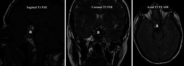
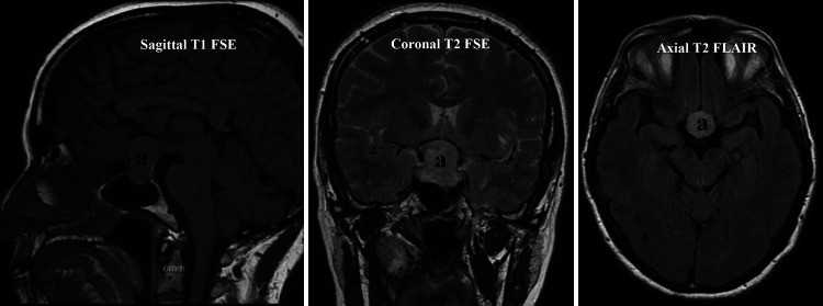
Observations: The case involves a 62-year-old female patient with a history of right carotid body tumor resection. She presented to the emergency department with a sudden decrease in visual acuity and bitemporal hemianopsia, accompanied by a left parietal headache. Initial brain magnetic resonance imaging (MRI) revealed a pituitary macroadenoma, which was completely resected. However, postoperatively, the patient developed left amaurosis. Subsequent brain MRI showed the presence of hemostatic material mixed with blood in the sellar region, causing displacement of the optic chiasm. A repeat intervention was performed, identifying bleeding from both cavernous sinuses. Head and neck angiography demonstrated a right glomus vagale tumor with abundant blood drainage into the right cavernous sinus. Embolization of the glomus vagale tumor was performed, resulting in no further bleeding and improvement of symptoms.
Lessons: The aim of this case report is to describe a rare occurrence of bilateral visual disturbances caused by bleeding in both cavernous sinuses due to venous hypertension caused by a right glomus vagale tumor.


 求助内容:
求助内容: 应助结果提醒方式:


