Ayman Hussien Shaamash, Mehad H AlQasem, Deama S Al Ghamdi, Ahmed A Mahfouz, Mamdoh A Eskandar
{"title":"仅通过MRI诊断的主要前置胎盘植入频谱:发病率、危险因素和母体发病率。","authors":"Ayman Hussien Shaamash, Mehad H AlQasem, Deama S Al Ghamdi, Ahmed A Mahfouz, Mamdoh A Eskandar","doi":"10.5144/0256-4947.2023.219","DOIUrl":null,"url":null,"abstract":"<p><strong>Background: </strong>Antenatal assessment of maternal risk factors and imaging evaluation can help in diagnosis and treatment of placenta accreta spectrum (PAS) in major placenta previa (PP). Recent evidence suggests that magnetic resonance imaging (MRI) could complement ultrasonography (US) in the PAS diagnosis.</p><p><strong>Objectives: </strong>Evaluate the incidence, risk factors, and maternal morbidity related to the MRI diagnosis of PAS in major PP.</p><p><strong>Design: </strong>A 10-year retrospective cohort study.</p><p><strong>Setting: </strong>Tertiary care hospital.</p><p><strong>Patients and methods: </strong>We report on patients with major PP who had cesarean delivery in Abha Maternity and Children's Hospital (AMCH) over a 10-year period (2012-2021). They were evaluated with ultrasonography (US) and color Doppler for evidence of PAS. Antenatal MRI was ordered either to confirm the diagnosis (if equivocal US) or to assess the depth of invasion/extra-uterine extension (if definitive US).</p><p><strong>Main outcome measures: </strong>Risk factors for PAS in major PP and maternal complications.</p><p><strong>Sample size: </strong>299 patients RESULTS: Among 299 patients, MRI confirmed the PAS diagnosis in 91/299 (30.5%) patients. The independent risk factors for MRI diagnosis of PAS in major PP included only repeated cesarean sections and advanced maternal age. The commonest maternal morbidity in major PP with PAS was significantly excessive intraoperative bleeding.</p><p><strong>Conclusion: </strong>MRI may be a valuable adjunct in the evaluation of PAS in major PP; as a complement, but not substitute US. MRI may be suitable in major PP/PAS patients who are older and have repeated cesarean deliveries with equivocal results or deep/extra-uterine extension on US.</p><p><strong>Limitation: </strong>Single center, small sample size, lack of complete histopathological diagnosis.</p><p><strong>Conflict of interest: </strong>None.</p>","PeriodicalId":8016,"journal":{"name":"Annals of Saudi Medicine","volume":"43 4","pages":"219-217"},"PeriodicalIF":1.8000,"publicationDate":"2023-07-01","publicationTypes":"Journal Article","fieldsOfStudy":null,"isOpenAccess":false,"openAccessPdf":"https://www.ncbi.nlm.nih.gov/pmc/articles/PMC10716833/pdf/","citationCount":"0","resultStr":"{\"title\":\"Placenta accreta spectrum in major placenta previa diagnosed only by MRI: incidence, risk factors, and maternal morbidity.\",\"authors\":\"Ayman Hussien Shaamash, Mehad H AlQasem, Deama S Al Ghamdi, Ahmed A Mahfouz, Mamdoh A Eskandar\",\"doi\":\"10.5144/0256-4947.2023.219\",\"DOIUrl\":null,\"url\":null,\"abstract\":\"<p><strong>Background: </strong>Antenatal assessment of maternal risk factors and imaging evaluation can help in diagnosis and treatment of placenta accreta spectrum (PAS) in major placenta previa (PP). Recent evidence suggests that magnetic resonance imaging (MRI) could complement ultrasonography (US) in the PAS diagnosis.</p><p><strong>Objectives: </strong>Evaluate the incidence, risk factors, and maternal morbidity related to the MRI diagnosis of PAS in major PP.</p><p><strong>Design: </strong>A 10-year retrospective cohort study.</p><p><strong>Setting: </strong>Tertiary care hospital.</p><p><strong>Patients and methods: </strong>We report on patients with major PP who had cesarean delivery in Abha Maternity and Children's Hospital (AMCH) over a 10-year period (2012-2021). They were evaluated with ultrasonography (US) and color Doppler for evidence of PAS. Antenatal MRI was ordered either to confirm the diagnosis (if equivocal US) or to assess the depth of invasion/extra-uterine extension (if definitive US).</p><p><strong>Main outcome measures: </strong>Risk factors for PAS in major PP and maternal complications.</p><p><strong>Sample size: </strong>299 patients RESULTS: Among 299 patients, MRI confirmed the PAS diagnosis in 91/299 (30.5%) patients. The independent risk factors for MRI diagnosis of PAS in major PP included only repeated cesarean sections and advanced maternal age. The commonest maternal morbidity in major PP with PAS was significantly excessive intraoperative bleeding.</p><p><strong>Conclusion: </strong>MRI may be a valuable adjunct in the evaluation of PAS in major PP; as a complement, but not substitute US. MRI may be suitable in major PP/PAS patients who are older and have repeated cesarean deliveries with equivocal results or deep/extra-uterine extension on US.</p><p><strong>Limitation: </strong>Single center, small sample size, lack of complete histopathological diagnosis.</p><p><strong>Conflict of interest: </strong>None.</p>\",\"PeriodicalId\":8016,\"journal\":{\"name\":\"Annals of Saudi Medicine\",\"volume\":\"43 4\",\"pages\":\"219-217\"},\"PeriodicalIF\":1.8000,\"publicationDate\":\"2023-07-01\",\"publicationTypes\":\"Journal Article\",\"fieldsOfStudy\":null,\"isOpenAccess\":false,\"openAccessPdf\":\"https://www.ncbi.nlm.nih.gov/pmc/articles/PMC10716833/pdf/\",\"citationCount\":\"0\",\"resultStr\":null,\"platform\":\"Semanticscholar\",\"paperid\":null,\"PeriodicalName\":\"Annals of Saudi Medicine\",\"FirstCategoryId\":\"3\",\"ListUrlMain\":\"https://doi.org/10.5144/0256-4947.2023.219\",\"RegionNum\":4,\"RegionCategory\":\"医学\",\"ArticlePicture\":[],\"TitleCN\":null,\"AbstractTextCN\":null,\"PMCID\":null,\"EPubDate\":\"2023/8/3 0:00:00\",\"PubModel\":\"Epub\",\"JCR\":\"Q2\",\"JCRName\":\"MEDICINE, GENERAL & INTERNAL\",\"Score\":null,\"Total\":0}","platform":"Semanticscholar","paperid":null,"PeriodicalName":"Annals of Saudi Medicine","FirstCategoryId":"3","ListUrlMain":"https://doi.org/10.5144/0256-4947.2023.219","RegionNum":4,"RegionCategory":"医学","ArticlePicture":[],"TitleCN":null,"AbstractTextCN":null,"PMCID":null,"EPubDate":"2023/8/3 0:00:00","PubModel":"Epub","JCR":"Q2","JCRName":"MEDICINE, GENERAL & INTERNAL","Score":null,"Total":0}
Placenta accreta spectrum in major placenta previa diagnosed only by MRI: incidence, risk factors, and maternal morbidity.
Background: Antenatal assessment of maternal risk factors and imaging evaluation can help in diagnosis and treatment of placenta accreta spectrum (PAS) in major placenta previa (PP). Recent evidence suggests that magnetic resonance imaging (MRI) could complement ultrasonography (US) in the PAS diagnosis.
Objectives: Evaluate the incidence, risk factors, and maternal morbidity related to the MRI diagnosis of PAS in major PP.
Design: A 10-year retrospective cohort study.
Setting: Tertiary care hospital.
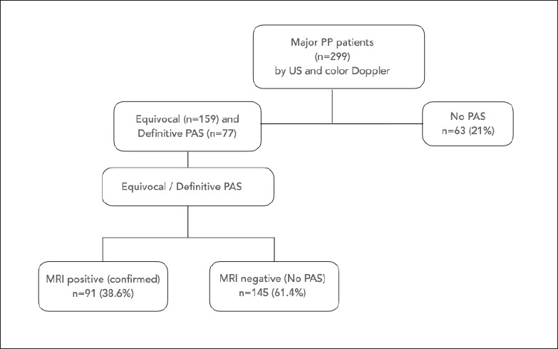
Patients and methods: We report on patients with major PP who had cesarean delivery in Abha Maternity and Children's Hospital (AMCH) over a 10-year period (2012-2021). They were evaluated with ultrasonography (US) and color Doppler for evidence of PAS. Antenatal MRI was ordered either to confirm the diagnosis (if equivocal US) or to assess the depth of invasion/extra-uterine extension (if definitive US).
Main outcome measures: Risk factors for PAS in major PP and maternal complications.
Sample size: 299 patients RESULTS: Among 299 patients, MRI confirmed the PAS diagnosis in 91/299 (30.5%) patients. The independent risk factors for MRI diagnosis of PAS in major PP included only repeated cesarean sections and advanced maternal age. The commonest maternal morbidity in major PP with PAS was significantly excessive intraoperative bleeding.
Conclusion: MRI may be a valuable adjunct in the evaluation of PAS in major PP; as a complement, but not substitute US. MRI may be suitable in major PP/PAS patients who are older and have repeated cesarean deliveries with equivocal results or deep/extra-uterine extension on US.
Limitation: Single center, small sample size, lack of complete histopathological diagnosis.
期刊介绍:
The Annals of Saudi Medicine (ASM) is published bimonthly by King Faisal Specialist Hospital and Research Centre, Riyadh, Saudi Arabia. We publish scientific reports of clinical interest in English. All submissions are subject to peer review by the editorial board and by reviewers in appropriate specialties. The journal will consider for publication manuscripts from any part of the world, but particularly reports that would be of interest to readers in the Middle East or other parts of Asia and Africa. Please go to the Author Resource Center for additional information.

 求助内容:
求助内容: 应助结果提醒方式:


