Ye Z Spector, Qi Zhao, Xiaopeng Zhao, William J Feuer, Portia Lynn Maravich, Xiang-Run Huang
{"title":"基于细胞骨架成分的轴突亚型分类。","authors":"Ye Z Spector, Qi Zhao, Xiaopeng Zhao, William J Feuer, Portia Lynn Maravich, Xiang-Run Huang","doi":"10.2147/CHC.S57081","DOIUrl":null,"url":null,"abstract":"<p><strong>Background: </strong>Retinal ganglion cells are often classified into different subtypes according to their morphology or physiological functions. The axons of RGCs contain three major cytoskeletal components: actin filaments (F-actin); microtubules; and neurofilaments (NFs). The contents of these components vary among axons. Our objective was to classify axons into subtypes based on the contents of cytoskeletal components and study their distributions across the retina in normal rodent retinas.</p><p><strong>Methods: </strong>Whole-mounted retinas of female Wistar rats were stained with phalloidin to label F-actin, anti-β-tubulin monoclonal antibody to mark microtubules, and antineurofilament antibody to label NFs. A confocal laser scanning microscope was used to provide en face images of retinal nerve fiber bundles with a resolution of 0.24 μm/pixel. Staining intensity profiles across axons were obtained for each cytoskeletal component. Axonal subtypes were then determined from the relative contents, indicated by the staining intensity, of these components. Linear density was used to investigate topographical distribution of each subtype across the retina.</p><p><strong>Results: </strong>Normal axons could be classified into seven subtypes - FMN, FM, FN, and MN subtypes, (in which at least two or three cytoskeletal components were intensely stained), and F, M, and N subtypes, (in which only one cytoskeletal component was intensely stained within an axon). The FMN subtype was the most abundant subtype. There was no preferential distribution of subtypes around the optic nerve head. However, the densities of the axonal subtypes that contained NFs were found significantly different in the central and peripheral retinal regions. Axonal sizes were subtype-dependent.</p><p><strong>Conclusion: </strong>Axons of retinal ganglion cells can be classified into different subtypes, based on the contents of axonal cytoskeletal components. The classified subtypes will provide a new means to study selective damage of axonal ultrastructures in ocular neuropathic diseases.</p>","PeriodicalId":72540,"journal":{"name":"Cell health and cytoskeleton","volume":"6 ","pages":"1-10"},"PeriodicalIF":0.0000,"publicationDate":"2014-01-01","publicationTypes":"Journal Article","fieldsOfStudy":null,"isOpenAccess":false,"openAccessPdf":"https://sci-hub-pdf.com/10.2147/CHC.S57081","citationCount":"1","resultStr":"{\"title\":\"Classification of axonal subtypes based on cytoskeletal components.\",\"authors\":\"Ye Z Spector, Qi Zhao, Xiaopeng Zhao, William J Feuer, Portia Lynn Maravich, Xiang-Run Huang\",\"doi\":\"10.2147/CHC.S57081\",\"DOIUrl\":null,\"url\":null,\"abstract\":\"<p><strong>Background: </strong>Retinal ganglion cells are often classified into different subtypes according to their morphology or physiological functions. The axons of RGCs contain three major cytoskeletal components: actin filaments (F-actin); microtubules; and neurofilaments (NFs). The contents of these components vary among axons. Our objective was to classify axons into subtypes based on the contents of cytoskeletal components and study their distributions across the retina in normal rodent retinas.</p><p><strong>Methods: </strong>Whole-mounted retinas of female Wistar rats were stained with phalloidin to label F-actin, anti-β-tubulin monoclonal antibody to mark microtubules, and antineurofilament antibody to label NFs. A confocal laser scanning microscope was used to provide en face images of retinal nerve fiber bundles with a resolution of 0.24 μm/pixel. Staining intensity profiles across axons were obtained for each cytoskeletal component. Axonal subtypes were then determined from the relative contents, indicated by the staining intensity, of these components. Linear density was used to investigate topographical distribution of each subtype across the retina.</p><p><strong>Results: </strong>Normal axons could be classified into seven subtypes - FMN, FM, FN, and MN subtypes, (in which at least two or three cytoskeletal components were intensely stained), and F, M, and N subtypes, (in which only one cytoskeletal component was intensely stained within an axon). The FMN subtype was the most abundant subtype. There was no preferential distribution of subtypes around the optic nerve head. However, the densities of the axonal subtypes that contained NFs were found significantly different in the central and peripheral retinal regions. Axonal sizes were subtype-dependent.</p><p><strong>Conclusion: </strong>Axons of retinal ganglion cells can be classified into different subtypes, based on the contents of axonal cytoskeletal components. The classified subtypes will provide a new means to study selective damage of axonal ultrastructures in ocular neuropathic diseases.</p>\",\"PeriodicalId\":72540,\"journal\":{\"name\":\"Cell health and cytoskeleton\",\"volume\":\"6 \",\"pages\":\"1-10\"},\"PeriodicalIF\":0.0000,\"publicationDate\":\"2014-01-01\",\"publicationTypes\":\"Journal Article\",\"fieldsOfStudy\":null,\"isOpenAccess\":false,\"openAccessPdf\":\"https://sci-hub-pdf.com/10.2147/CHC.S57081\",\"citationCount\":\"1\",\"resultStr\":null,\"platform\":\"Semanticscholar\",\"paperid\":null,\"PeriodicalName\":\"Cell health and cytoskeleton\",\"FirstCategoryId\":\"1085\",\"ListUrlMain\":\"https://doi.org/10.2147/CHC.S57081\",\"RegionNum\":0,\"RegionCategory\":null,\"ArticlePicture\":[],\"TitleCN\":null,\"AbstractTextCN\":null,\"PMCID\":null,\"EPubDate\":\"\",\"PubModel\":\"\",\"JCR\":\"\",\"JCRName\":\"\",\"Score\":null,\"Total\":0}","platform":"Semanticscholar","paperid":null,"PeriodicalName":"Cell health and cytoskeleton","FirstCategoryId":"1085","ListUrlMain":"https://doi.org/10.2147/CHC.S57081","RegionNum":0,"RegionCategory":null,"ArticlePicture":[],"TitleCN":null,"AbstractTextCN":null,"PMCID":null,"EPubDate":"","PubModel":"","JCR":"","JCRName":"","Score":null,"Total":0}
Classification of axonal subtypes based on cytoskeletal components.
Background: Retinal ganglion cells are often classified into different subtypes according to their morphology or physiological functions. The axons of RGCs contain three major cytoskeletal components: actin filaments (F-actin); microtubules; and neurofilaments (NFs). The contents of these components vary among axons. Our objective was to classify axons into subtypes based on the contents of cytoskeletal components and study their distributions across the retina in normal rodent retinas.
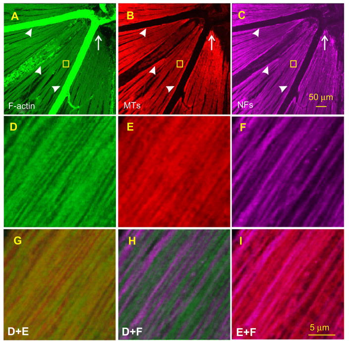
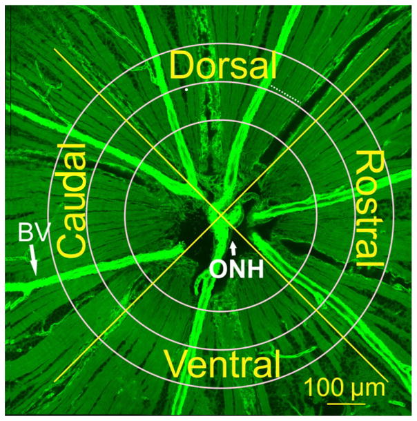
Methods: Whole-mounted retinas of female Wistar rats were stained with phalloidin to label F-actin, anti-β-tubulin monoclonal antibody to mark microtubules, and antineurofilament antibody to label NFs. A confocal laser scanning microscope was used to provide en face images of retinal nerve fiber bundles with a resolution of 0.24 μm/pixel. Staining intensity profiles across axons were obtained for each cytoskeletal component. Axonal subtypes were then determined from the relative contents, indicated by the staining intensity, of these components. Linear density was used to investigate topographical distribution of each subtype across the retina.
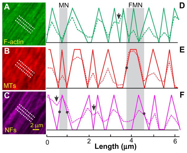
Results: Normal axons could be classified into seven subtypes - FMN, FM, FN, and MN subtypes, (in which at least two or three cytoskeletal components were intensely stained), and F, M, and N subtypes, (in which only one cytoskeletal component was intensely stained within an axon). The FMN subtype was the most abundant subtype. There was no preferential distribution of subtypes around the optic nerve head. However, the densities of the axonal subtypes that contained NFs were found significantly different in the central and peripheral retinal regions. Axonal sizes were subtype-dependent.
Conclusion: Axons of retinal ganglion cells can be classified into different subtypes, based on the contents of axonal cytoskeletal components. The classified subtypes will provide a new means to study selective damage of axonal ultrastructures in ocular neuropathic diseases.

 求助内容:
求助内容: 应助结果提醒方式:


