{"title":"特殊类型食管癌的临床预测因素。","authors":"Yugo Suzuki, Yu Ohkura, Mako Koseki, Kosuke Nomura, Akira Matsui, Masaki Ueno, Daisuke Kikuchi, Kenichi Ohashi, Shu Hoteya","doi":"10.1007/s10388-023-01003-1","DOIUrl":null,"url":null,"abstract":"<p><strong>Background: </strong>Esophageal cancers with a histological type other than the two major types, squamous cell carcinoma (SCC) and adenocarcinoma, are referred to as \"special type of esophageal cancer\" (STEC). STEC is rare and difficult to diagnose preoperatively. Therefore, we aimed to clarify the clinicopathological findings of STEC, including magnifying endoscopy with narrow band imaging (ME-NBI).</p><p><strong>Methods: </strong>We reviewed 1133 lesions in 936 consecutive cases who underwent endoscopic resection or surgical resection for primary esophageal cancer. Patients were classified into the SCC group and the STEC group, respectively. Factors that predict STEC endoscopically, as well as clinicopathologic features of STEC compared to SCC, were examined.</p><p><strong>Results: </strong>Twenty-eight STECs were diagnosed in 28 patients: 15 with basaloid squamous cell carcinoma, 6 with adenosquamous carcinoma, 4 with mucoepidermoid carcinoma, 1 with carcinosarcoma, 1 with salivary duct-type carcinoma, and 1 with neuroendocrine cell carcinoma. There was significantly more pT1b or deeper cancer (60.7% vs. 12.8%), lymphovascular invasion (50.0% vs. 11.1%) and elevated type (53.6% vs. 16.1%) in the STEC group. The proportion of lesions with type R vessels on ME-NBI was significantly higher in the STEC group (46.4% vs. 3.9%). The STEC group had significantly lower accuracy of ME-NBI for prediction of depth (64.3% vs. 83.5%) and a greater proportion of underestimated lesions (32.1% vs. 9.3%). In the multivariate analysis, the histopathology of STEC was associated with type R vessels on ME-NBI.</p><p><strong>Conclusion: </strong>Type R vessels and submucosal tumor-like elevation might be the clinical predictors of STEC.</p>","PeriodicalId":11918,"journal":{"name":"Esophagus","volume":null,"pages":null},"PeriodicalIF":2.2000,"publicationDate":"2023-07-01","publicationTypes":"Journal Article","fieldsOfStudy":null,"isOpenAccess":false,"openAccessPdf":"","citationCount":"0","resultStr":"{\"title\":\"Clinical predictors of special type of esophageal cancer.\",\"authors\":\"Yugo Suzuki, Yu Ohkura, Mako Koseki, Kosuke Nomura, Akira Matsui, Masaki Ueno, Daisuke Kikuchi, Kenichi Ohashi, Shu Hoteya\",\"doi\":\"10.1007/s10388-023-01003-1\",\"DOIUrl\":null,\"url\":null,\"abstract\":\"<p><strong>Background: </strong>Esophageal cancers with a histological type other than the two major types, squamous cell carcinoma (SCC) and adenocarcinoma, are referred to as \\\"special type of esophageal cancer\\\" (STEC). STEC is rare and difficult to diagnose preoperatively. Therefore, we aimed to clarify the clinicopathological findings of STEC, including magnifying endoscopy with narrow band imaging (ME-NBI).</p><p><strong>Methods: </strong>We reviewed 1133 lesions in 936 consecutive cases who underwent endoscopic resection or surgical resection for primary esophageal cancer. Patients were classified into the SCC group and the STEC group, respectively. Factors that predict STEC endoscopically, as well as clinicopathologic features of STEC compared to SCC, were examined.</p><p><strong>Results: </strong>Twenty-eight STECs were diagnosed in 28 patients: 15 with basaloid squamous cell carcinoma, 6 with adenosquamous carcinoma, 4 with mucoepidermoid carcinoma, 1 with carcinosarcoma, 1 with salivary duct-type carcinoma, and 1 with neuroendocrine cell carcinoma. There was significantly more pT1b or deeper cancer (60.7% vs. 12.8%), lymphovascular invasion (50.0% vs. 11.1%) and elevated type (53.6% vs. 16.1%) in the STEC group. The proportion of lesions with type R vessels on ME-NBI was significantly higher in the STEC group (46.4% vs. 3.9%). The STEC group had significantly lower accuracy of ME-NBI for prediction of depth (64.3% vs. 83.5%) and a greater proportion of underestimated lesions (32.1% vs. 9.3%). In the multivariate analysis, the histopathology of STEC was associated with type R vessels on ME-NBI.</p><p><strong>Conclusion: </strong>Type R vessels and submucosal tumor-like elevation might be the clinical predictors of STEC.</p>\",\"PeriodicalId\":11918,\"journal\":{\"name\":\"Esophagus\",\"volume\":null,\"pages\":null},\"PeriodicalIF\":2.2000,\"publicationDate\":\"2023-07-01\",\"publicationTypes\":\"Journal Article\",\"fieldsOfStudy\":null,\"isOpenAccess\":false,\"openAccessPdf\":\"\",\"citationCount\":\"0\",\"resultStr\":null,\"platform\":\"Semanticscholar\",\"paperid\":null,\"PeriodicalName\":\"Esophagus\",\"FirstCategoryId\":\"3\",\"ListUrlMain\":\"https://doi.org/10.1007/s10388-023-01003-1\",\"RegionNum\":4,\"RegionCategory\":\"医学\",\"ArticlePicture\":[],\"TitleCN\":null,\"AbstractTextCN\":null,\"PMCID\":null,\"EPubDate\":\"\",\"PubModel\":\"\",\"JCR\":\"Q3\",\"JCRName\":\"GASTROENTEROLOGY & HEPATOLOGY\",\"Score\":null,\"Total\":0}","platform":"Semanticscholar","paperid":null,"PeriodicalName":"Esophagus","FirstCategoryId":"3","ListUrlMain":"https://doi.org/10.1007/s10388-023-01003-1","RegionNum":4,"RegionCategory":"医学","ArticlePicture":[],"TitleCN":null,"AbstractTextCN":null,"PMCID":null,"EPubDate":"","PubModel":"","JCR":"Q3","JCRName":"GASTROENTEROLOGY & HEPATOLOGY","Score":null,"Total":0}
Clinical predictors of special type of esophageal cancer.
Background: Esophageal cancers with a histological type other than the two major types, squamous cell carcinoma (SCC) and adenocarcinoma, are referred to as "special type of esophageal cancer" (STEC). STEC is rare and difficult to diagnose preoperatively. Therefore, we aimed to clarify the clinicopathological findings of STEC, including magnifying endoscopy with narrow band imaging (ME-NBI).
Methods: We reviewed 1133 lesions in 936 consecutive cases who underwent endoscopic resection or surgical resection for primary esophageal cancer. Patients were classified into the SCC group and the STEC group, respectively. Factors that predict STEC endoscopically, as well as clinicopathologic features of STEC compared to SCC, were examined.
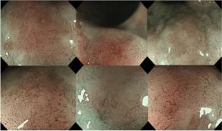
Results: Twenty-eight STECs were diagnosed in 28 patients: 15 with basaloid squamous cell carcinoma, 6 with adenosquamous carcinoma, 4 with mucoepidermoid carcinoma, 1 with carcinosarcoma, 1 with salivary duct-type carcinoma, and 1 with neuroendocrine cell carcinoma. There was significantly more pT1b or deeper cancer (60.7% vs. 12.8%), lymphovascular invasion (50.0% vs. 11.1%) and elevated type (53.6% vs. 16.1%) in the STEC group. The proportion of lesions with type R vessels on ME-NBI was significantly higher in the STEC group (46.4% vs. 3.9%). The STEC group had significantly lower accuracy of ME-NBI for prediction of depth (64.3% vs. 83.5%) and a greater proportion of underestimated lesions (32.1% vs. 9.3%). In the multivariate analysis, the histopathology of STEC was associated with type R vessels on ME-NBI.
Conclusion: Type R vessels and submucosal tumor-like elevation might be the clinical predictors of STEC.
期刊介绍:
Esophagus, the official journal of the Japan Esophageal Society, introduces practitioners and researchers to significant studies in the fields of benign and malignant diseases of the esophagus. The journal welcomes original articles, review articles, and short articles including technical notes ( How I do it ), which will be peer-reviewed by the editorial board. Letters to the editor are also welcome. Special articles on esophageal diseases will be provided by the editorial board, and proceedings of symposia and workshops will be included in special issues for the Annual Congress of the Society.
 求助内容:
求助内容: 应助结果提醒方式:


