Amarit Kay Gill, Ashok Raghavan, Eishaan Kamta Bhargava
{"title":"孤立性先天性面神经发育不全。","authors":"Amarit Kay Gill, Ashok Raghavan, Eishaan Kamta Bhargava","doi":"10.1259/bjrcr.20220119","DOIUrl":null,"url":null,"abstract":"<p><p>An otherwise healthy 2-month-old boy was referred to ENT for a congenital right facial palsy, with a birth history of difficult ventouse delivery. Initially, a traumatic cause was suspected, however subsequent MR 3D-FIESTA (<i>T<sub>2</sub></i> weighted) imaging demonstrated a right facial nerve agenesis with normal appearances of the remainder of the brain parenchyma, cranial nerves and parotid glands. There were no syndromic features or hearing difficulties. Isolated congenital nerve agenesis is a rare condition, with very few case reports available in the literature. Pre-natal 4D ultrasound imaging further supports the diagnosis. To our knowledge, this is the first published pre-natal ultrasound image of congenital facial nerve palsy. The infant has been referred for consideration of nerve reconstruction surgery, and is receiving multi-disciplinary input from ENT, Physiotherapy and Ophthalmology, the latter for prevention of exposure keratitis.</p>","PeriodicalId":45216,"journal":{"name":"BJR Case Reports","volume":"9 3","pages":"20220119"},"PeriodicalIF":0.5000,"publicationDate":"2023-05-01","publicationTypes":"Journal Article","fieldsOfStudy":null,"isOpenAccess":false,"openAccessPdf":"https://www.ncbi.nlm.nih.gov/pmc/articles/PMC10230233/pdf/","citationCount":"0","resultStr":"{\"title\":\"Isolated congenital facial nerve agenesis.\",\"authors\":\"Amarit Kay Gill, Ashok Raghavan, Eishaan Kamta Bhargava\",\"doi\":\"10.1259/bjrcr.20220119\",\"DOIUrl\":null,\"url\":null,\"abstract\":\"<p><p>An otherwise healthy 2-month-old boy was referred to ENT for a congenital right facial palsy, with a birth history of difficult ventouse delivery. Initially, a traumatic cause was suspected, however subsequent MR 3D-FIESTA (<i>T<sub>2</sub></i> weighted) imaging demonstrated a right facial nerve agenesis with normal appearances of the remainder of the brain parenchyma, cranial nerves and parotid glands. There were no syndromic features or hearing difficulties. Isolated congenital nerve agenesis is a rare condition, with very few case reports available in the literature. Pre-natal 4D ultrasound imaging further supports the diagnosis. To our knowledge, this is the first published pre-natal ultrasound image of congenital facial nerve palsy. The infant has been referred for consideration of nerve reconstruction surgery, and is receiving multi-disciplinary input from ENT, Physiotherapy and Ophthalmology, the latter for prevention of exposure keratitis.</p>\",\"PeriodicalId\":45216,\"journal\":{\"name\":\"BJR Case Reports\",\"volume\":\"9 3\",\"pages\":\"20220119\"},\"PeriodicalIF\":0.5000,\"publicationDate\":\"2023-05-01\",\"publicationTypes\":\"Journal Article\",\"fieldsOfStudy\":null,\"isOpenAccess\":false,\"openAccessPdf\":\"https://www.ncbi.nlm.nih.gov/pmc/articles/PMC10230233/pdf/\",\"citationCount\":\"0\",\"resultStr\":null,\"platform\":\"Semanticscholar\",\"paperid\":null,\"PeriodicalName\":\"BJR Case Reports\",\"FirstCategoryId\":\"1085\",\"ListUrlMain\":\"https://doi.org/10.1259/bjrcr.20220119\",\"RegionNum\":0,\"RegionCategory\":null,\"ArticlePicture\":[],\"TitleCN\":null,\"AbstractTextCN\":null,\"PMCID\":null,\"EPubDate\":\"\",\"PubModel\":\"\",\"JCR\":\"Q4\",\"JCRName\":\"RADIOLOGY, NUCLEAR MEDICINE & MEDICAL IMAGING\",\"Score\":null,\"Total\":0}","platform":"Semanticscholar","paperid":null,"PeriodicalName":"BJR Case Reports","FirstCategoryId":"1085","ListUrlMain":"https://doi.org/10.1259/bjrcr.20220119","RegionNum":0,"RegionCategory":null,"ArticlePicture":[],"TitleCN":null,"AbstractTextCN":null,"PMCID":null,"EPubDate":"","PubModel":"","JCR":"Q4","JCRName":"RADIOLOGY, NUCLEAR MEDICINE & MEDICAL IMAGING","Score":null,"Total":0}
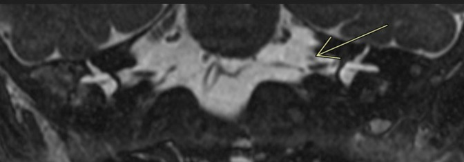
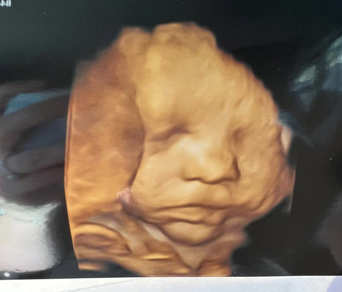
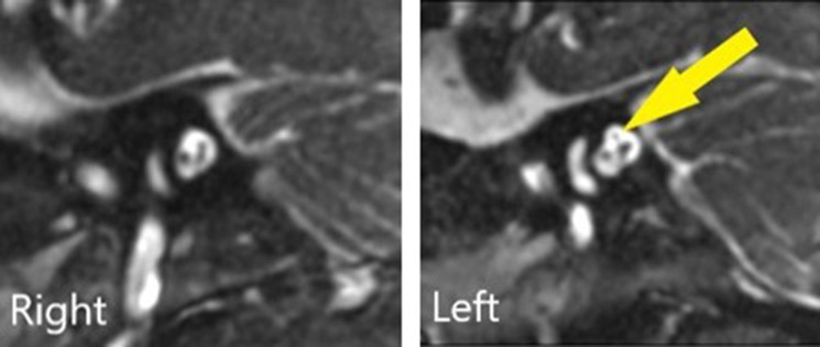
An otherwise healthy 2-month-old boy was referred to ENT for a congenital right facial palsy, with a birth history of difficult ventouse delivery. Initially, a traumatic cause was suspected, however subsequent MR 3D-FIESTA (T2 weighted) imaging demonstrated a right facial nerve agenesis with normal appearances of the remainder of the brain parenchyma, cranial nerves and parotid glands. There were no syndromic features or hearing difficulties. Isolated congenital nerve agenesis is a rare condition, with very few case reports available in the literature. Pre-natal 4D ultrasound imaging further supports the diagnosis. To our knowledge, this is the first published pre-natal ultrasound image of congenital facial nerve palsy. The infant has been referred for consideration of nerve reconstruction surgery, and is receiving multi-disciplinary input from ENT, Physiotherapy and Ophthalmology, the latter for prevention of exposure keratitis.

 求助内容:
求助内容: 应助结果提醒方式:


