Direnç Özlem Aksoy, Yeşim Karagöz, Kemal Furkan Kaldırımoğlu, Melis Baykara Ulusan, Abdullah Soydan Mahmutoğlu
{"title":"听觉通路的扩散张量成像:儿童耳蜗植入物候选者和健康病例的比较。","authors":"Direnç Özlem Aksoy, Yeşim Karagöz, Kemal Furkan Kaldırımoğlu, Melis Baykara Ulusan, Abdullah Soydan Mahmutoğlu","doi":"10.5152/iao.2023.22998","DOIUrl":null,"url":null,"abstract":"<p><strong>Background: </strong>We aimed to investigate the changes that may occur in the auditory neural network in pediatric congenital hearing loss cases.</p><p><strong>Methods: </strong>Fifty-four cochlear implant candidates and 47 normal-hearing controls were included in this retrospective study. Fractional anisotropy, radial diffusivity, and apparent diffusion coefficient maps were generated. We placed region of interest on the cochlear nucleus, superior olivary nucleus, lateral lemniscus, medial geniculate body, auditory radiation, Heschl's gyrus, inferior fronto-occipital fasciculus, superior longitudinal fascicle, and corpus callosum splenium. The area of the cochlear nerve was measured. Diffusion tensor imaging metrics, children's ages, and cochlear nerve area were compared.</p><p><strong>Results: </strong>Apparent diffusion coefficient and radial diffusivity values of patients were higher than the control group in all places except the radial diffusivity values of medial geniculate body. The fractional anisotropy values of the patients in lateral lemniscus, auditory radiation, Heschl's gyrus, inferior fronto-occipital fasciculus, superior longitudinal fascicle, and corpus callosum splenium were lower than the control group. There is a positive correlation between fractional anisotropy and age in both patient and control groups for all locations. The cochlear nerve area is lower in patients (0.88 ± 0.29) than in the control group (1.18 ± 0.14) (P = .000). The cochlear nerve area has a positive correlation with age in the patient group (P = .000) but has not in the control group. The cochlear nerve area positively correlates with fractional anisotropy values of all locations except fractional anisotropy values of medial geniculate body.</p><p><strong>Conclusion: </strong>The alterations of diffusion tensor imaging metrics on the auditory pathway reflect the microstructural changes of white matter tracts.</p>","PeriodicalId":54793,"journal":{"name":"Journal of International Advanced Otology","volume":"19 4","pages":"333-341"},"PeriodicalIF":1.0000,"publicationDate":"2023-07-01","publicationTypes":"Journal Article","fieldsOfStudy":null,"isOpenAccess":false,"openAccessPdf":"https://ftp.ncbi.nlm.nih.gov/pub/pmc/oa_pdf/2f/bb/jiao-19-4-333.PMC10544541.pdf","citationCount":"0","resultStr":"{\"title\":\"Diffusion Tensor Imaging of Auditory Pathway: A Comparison of Pediatric Cochlear Implant Candidates and Healthy Cases.\",\"authors\":\"Direnç Özlem Aksoy, Yeşim Karagöz, Kemal Furkan Kaldırımoğlu, Melis Baykara Ulusan, Abdullah Soydan Mahmutoğlu\",\"doi\":\"10.5152/iao.2023.22998\",\"DOIUrl\":null,\"url\":null,\"abstract\":\"<p><strong>Background: </strong>We aimed to investigate the changes that may occur in the auditory neural network in pediatric congenital hearing loss cases.</p><p><strong>Methods: </strong>Fifty-four cochlear implant candidates and 47 normal-hearing controls were included in this retrospective study. Fractional anisotropy, radial diffusivity, and apparent diffusion coefficient maps were generated. We placed region of interest on the cochlear nucleus, superior olivary nucleus, lateral lemniscus, medial geniculate body, auditory radiation, Heschl's gyrus, inferior fronto-occipital fasciculus, superior longitudinal fascicle, and corpus callosum splenium. The area of the cochlear nerve was measured. Diffusion tensor imaging metrics, children's ages, and cochlear nerve area were compared.</p><p><strong>Results: </strong>Apparent diffusion coefficient and radial diffusivity values of patients were higher than the control group in all places except the radial diffusivity values of medial geniculate body. The fractional anisotropy values of the patients in lateral lemniscus, auditory radiation, Heschl's gyrus, inferior fronto-occipital fasciculus, superior longitudinal fascicle, and corpus callosum splenium were lower than the control group. There is a positive correlation between fractional anisotropy and age in both patient and control groups for all locations. The cochlear nerve area is lower in patients (0.88 ± 0.29) than in the control group (1.18 ± 0.14) (P = .000). The cochlear nerve area has a positive correlation with age in the patient group (P = .000) but has not in the control group. The cochlear nerve area positively correlates with fractional anisotropy values of all locations except fractional anisotropy values of medial geniculate body.</p><p><strong>Conclusion: </strong>The alterations of diffusion tensor imaging metrics on the auditory pathway reflect the microstructural changes of white matter tracts.</p>\",\"PeriodicalId\":54793,\"journal\":{\"name\":\"Journal of International Advanced Otology\",\"volume\":\"19 4\",\"pages\":\"333-341\"},\"PeriodicalIF\":1.0000,\"publicationDate\":\"2023-07-01\",\"publicationTypes\":\"Journal Article\",\"fieldsOfStudy\":null,\"isOpenAccess\":false,\"openAccessPdf\":\"https://ftp.ncbi.nlm.nih.gov/pub/pmc/oa_pdf/2f/bb/jiao-19-4-333.PMC10544541.pdf\",\"citationCount\":\"0\",\"resultStr\":null,\"platform\":\"Semanticscholar\",\"paperid\":null,\"PeriodicalName\":\"Journal of International Advanced Otology\",\"FirstCategoryId\":\"3\",\"ListUrlMain\":\"https://doi.org/10.5152/iao.2023.22998\",\"RegionNum\":4,\"RegionCategory\":\"医学\",\"ArticlePicture\":[],\"TitleCN\":null,\"AbstractTextCN\":null,\"PMCID\":null,\"EPubDate\":\"\",\"PubModel\":\"\",\"JCR\":\"Q3\",\"JCRName\":\"OTORHINOLARYNGOLOGY\",\"Score\":null,\"Total\":0}","platform":"Semanticscholar","paperid":null,"PeriodicalName":"Journal of International Advanced Otology","FirstCategoryId":"3","ListUrlMain":"https://doi.org/10.5152/iao.2023.22998","RegionNum":4,"RegionCategory":"医学","ArticlePicture":[],"TitleCN":null,"AbstractTextCN":null,"PMCID":null,"EPubDate":"","PubModel":"","JCR":"Q3","JCRName":"OTORHINOLARYNGOLOGY","Score":null,"Total":0}
Diffusion Tensor Imaging of Auditory Pathway: A Comparison of Pediatric Cochlear Implant Candidates and Healthy Cases.
Background: We aimed to investigate the changes that may occur in the auditory neural network in pediatric congenital hearing loss cases.
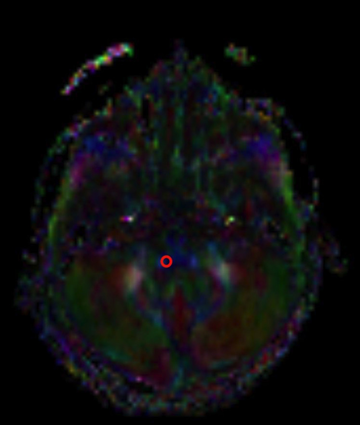
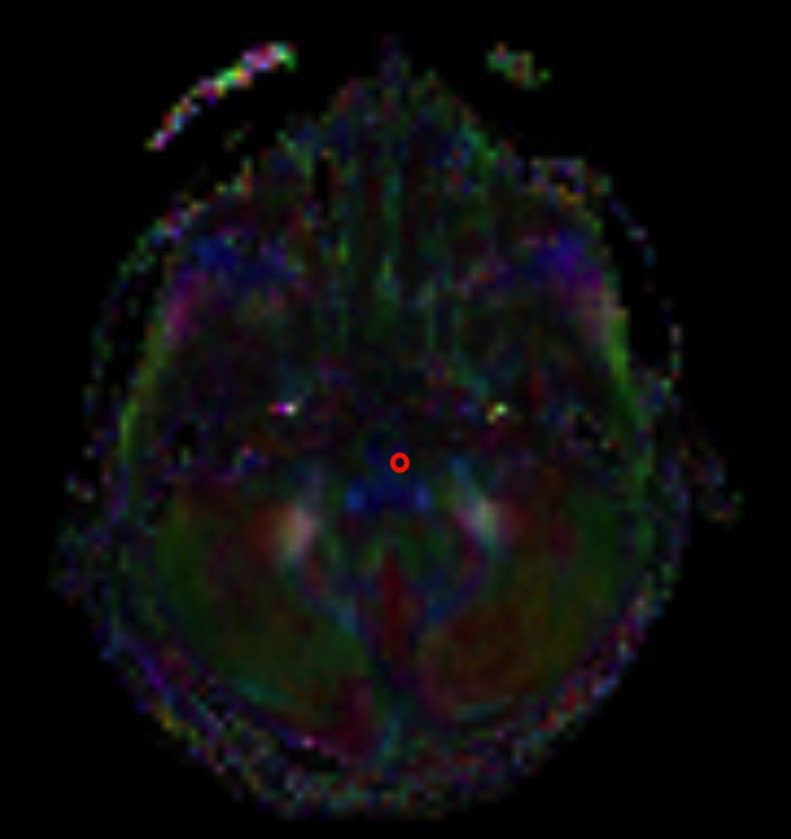
Methods: Fifty-four cochlear implant candidates and 47 normal-hearing controls were included in this retrospective study. Fractional anisotropy, radial diffusivity, and apparent diffusion coefficient maps were generated. We placed region of interest on the cochlear nucleus, superior olivary nucleus, lateral lemniscus, medial geniculate body, auditory radiation, Heschl's gyrus, inferior fronto-occipital fasciculus, superior longitudinal fascicle, and corpus callosum splenium. The area of the cochlear nerve was measured. Diffusion tensor imaging metrics, children's ages, and cochlear nerve area were compared.
Results: Apparent diffusion coefficient and radial diffusivity values of patients were higher than the control group in all places except the radial diffusivity values of medial geniculate body. The fractional anisotropy values of the patients in lateral lemniscus, auditory radiation, Heschl's gyrus, inferior fronto-occipital fasciculus, superior longitudinal fascicle, and corpus callosum splenium were lower than the control group. There is a positive correlation between fractional anisotropy and age in both patient and control groups for all locations. The cochlear nerve area is lower in patients (0.88 ± 0.29) than in the control group (1.18 ± 0.14) (P = .000). The cochlear nerve area has a positive correlation with age in the patient group (P = .000) but has not in the control group. The cochlear nerve area positively correlates with fractional anisotropy values of all locations except fractional anisotropy values of medial geniculate body.
Conclusion: The alterations of diffusion tensor imaging metrics on the auditory pathway reflect the microstructural changes of white matter tracts.
期刊介绍:
The Journal of International Advanced Otology (IAO – Citation Abbreviation: J Int Adv Otol) is an open access double-blind peer-reviewed, international publication. The Journal of International Advanced Otology is fully sponsored and owned by the European Academy of Otology and Neurotology and the Politzer Society. The Journal of International Advanced Otology is published 3 times per year on April, August, December and its publication language is English.
The scope of the Journal is limited with otology, neurotology, audiology (excluding linguistics) and skull base medicine.
The Journal of International Advanced Otology aims to publish manuscripts at the highest clinical and scientific level. IAO publishes original articles in the form of clinical and basic research, review articles, short reports and a limited number of case reports. Controversial patient discussions, communications on emerging technology, and historical issues will also be considered for publication.


 求助内容:
求助内容: 应助结果提醒方式:


