Asha Krishnakumar, Ashwin Ghadiyaram, Vishal C Patel, Charles F Opalak, Neel Dixit, William C Broaddus
{"title":"浅表性铁质沉着症:两个病例的比较表明有两个不同的诊断实体。示例性案例。","authors":"Asha Krishnakumar, Ashwin Ghadiyaram, Vishal C Patel, Charles F Opalak, Neel Dixit, William C Broaddus","doi":"10.3171/CASE23161","DOIUrl":null,"url":null,"abstract":"<p><strong>Background: </strong>Superficial siderosis is the deposition of hemosiderin in the superficial layers of the central nervous system. It has been described in patients with chronic leakage of blood into the cerebrospinal fluid or with amyloid angiopathy, often associated with Alzheimer's disease (AD).</p><p><strong>Observations: </strong>We present two cases of superficial siderosis with vastly different symptomatologies and treatment courses. The patient in case 1 had diffuse superficial siderosis demonstrated on T2-weighted magnetic resonance imaging (MRI), appearing mostly in the inferior cerebellum and extending throughout the neuraxis. He presented with hearing loss, spasticity, gait abnormalities, and urinary incontinence. Ultimately, surgical exploration of the thoracic spinal dura revealed an arteriovenous fistula, which was obliterated. His clinical course stabilized but with persistent deficits. The patient in case 2 had a family history of AD and underwent MRI to evaluate for memory impairment, which demonstrated superficial siderosis of the left occipital lobe. Lumbar puncture demonstrated only traumatic contamination by red blood cells, but tau protein analysis was consistent with the diagnosis of AD.</p><p><strong>Lessons: </strong>Superficial siderosis is a diagnostic term prompted by findings on MRI that can arise due to two different pathological entities. The diagnosis in case 1 should be termed diffuse superficial siderosis and in case 2 should be termed lobar cortical siderosis.</p>","PeriodicalId":16554,"journal":{"name":"Journal of Neurosurgery: Case Lessons","volume":"5 22","pages":""},"PeriodicalIF":0.0000,"publicationDate":"2023-05-29","publicationTypes":"Journal Article","fieldsOfStudy":null,"isOpenAccess":false,"openAccessPdf":"https://ftp.ncbi.nlm.nih.gov/pub/pmc/oa_pdf/23/b9/CASE23161.PMC10550672.pdf","citationCount":"0","resultStr":"{\"title\":\"Superficial siderosis: comparison of two cases indicates two distinct diagnostic entities. Illustrative cases.\",\"authors\":\"Asha Krishnakumar, Ashwin Ghadiyaram, Vishal C Patel, Charles F Opalak, Neel Dixit, William C Broaddus\",\"doi\":\"10.3171/CASE23161\",\"DOIUrl\":null,\"url\":null,\"abstract\":\"<p><strong>Background: </strong>Superficial siderosis is the deposition of hemosiderin in the superficial layers of the central nervous system. It has been described in patients with chronic leakage of blood into the cerebrospinal fluid or with amyloid angiopathy, often associated with Alzheimer's disease (AD).</p><p><strong>Observations: </strong>We present two cases of superficial siderosis with vastly different symptomatologies and treatment courses. The patient in case 1 had diffuse superficial siderosis demonstrated on T2-weighted magnetic resonance imaging (MRI), appearing mostly in the inferior cerebellum and extending throughout the neuraxis. He presented with hearing loss, spasticity, gait abnormalities, and urinary incontinence. Ultimately, surgical exploration of the thoracic spinal dura revealed an arteriovenous fistula, which was obliterated. His clinical course stabilized but with persistent deficits. The patient in case 2 had a family history of AD and underwent MRI to evaluate for memory impairment, which demonstrated superficial siderosis of the left occipital lobe. Lumbar puncture demonstrated only traumatic contamination by red blood cells, but tau protein analysis was consistent with the diagnosis of AD.</p><p><strong>Lessons: </strong>Superficial siderosis is a diagnostic term prompted by findings on MRI that can arise due to two different pathological entities. The diagnosis in case 1 should be termed diffuse superficial siderosis and in case 2 should be termed lobar cortical siderosis.</p>\",\"PeriodicalId\":16554,\"journal\":{\"name\":\"Journal of Neurosurgery: Case Lessons\",\"volume\":\"5 22\",\"pages\":\"\"},\"PeriodicalIF\":0.0000,\"publicationDate\":\"2023-05-29\",\"publicationTypes\":\"Journal Article\",\"fieldsOfStudy\":null,\"isOpenAccess\":false,\"openAccessPdf\":\"https://ftp.ncbi.nlm.nih.gov/pub/pmc/oa_pdf/23/b9/CASE23161.PMC10550672.pdf\",\"citationCount\":\"0\",\"resultStr\":null,\"platform\":\"Semanticscholar\",\"paperid\":null,\"PeriodicalName\":\"Journal of Neurosurgery: Case Lessons\",\"FirstCategoryId\":\"1085\",\"ListUrlMain\":\"https://doi.org/10.3171/CASE23161\",\"RegionNum\":0,\"RegionCategory\":null,\"ArticlePicture\":[],\"TitleCN\":null,\"AbstractTextCN\":null,\"PMCID\":null,\"EPubDate\":\"\",\"PubModel\":\"\",\"JCR\":\"\",\"JCRName\":\"\",\"Score\":null,\"Total\":0}","platform":"Semanticscholar","paperid":null,"PeriodicalName":"Journal of Neurosurgery: Case Lessons","FirstCategoryId":"1085","ListUrlMain":"https://doi.org/10.3171/CASE23161","RegionNum":0,"RegionCategory":null,"ArticlePicture":[],"TitleCN":null,"AbstractTextCN":null,"PMCID":null,"EPubDate":"","PubModel":"","JCR":"","JCRName":"","Score":null,"Total":0}
Superficial siderosis: comparison of two cases indicates two distinct diagnostic entities. Illustrative cases.
Background: Superficial siderosis is the deposition of hemosiderin in the superficial layers of the central nervous system. It has been described in patients with chronic leakage of blood into the cerebrospinal fluid or with amyloid angiopathy, often associated with Alzheimer's disease (AD).
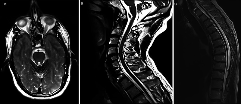
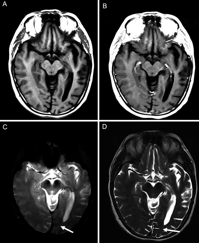
Observations: We present two cases of superficial siderosis with vastly different symptomatologies and treatment courses. The patient in case 1 had diffuse superficial siderosis demonstrated on T2-weighted magnetic resonance imaging (MRI), appearing mostly in the inferior cerebellum and extending throughout the neuraxis. He presented with hearing loss, spasticity, gait abnormalities, and urinary incontinence. Ultimately, surgical exploration of the thoracic spinal dura revealed an arteriovenous fistula, which was obliterated. His clinical course stabilized but with persistent deficits. The patient in case 2 had a family history of AD and underwent MRI to evaluate for memory impairment, which demonstrated superficial siderosis of the left occipital lobe. Lumbar puncture demonstrated only traumatic contamination by red blood cells, but tau protein analysis was consistent with the diagnosis of AD.
Lessons: Superficial siderosis is a diagnostic term prompted by findings on MRI that can arise due to two different pathological entities. The diagnosis in case 1 should be termed diffuse superficial siderosis and in case 2 should be termed lobar cortical siderosis.

 求助内容:
求助内容: 应助结果提醒方式:


