John P. Bois MD , Chadi Ayoub MBBS, PhD , Jeffrey B. Geske MD , Yee Weng Wong MBBS, MHS , Muhannad A. Abbasi MBBCh , Thomas A. Foley MD , Sharon L. Mulvagh MD , Christopher G. Scott MS , Steve R. Ommen MD , Patricia A. Pellikka MD
{"title":"超声增强剂与经胸超声心动图对肥厚性心肌病最大壁厚的测定","authors":"John P. Bois MD , Chadi Ayoub MBBS, PhD , Jeffrey B. Geske MD , Yee Weng Wong MBBS, MHS , Muhannad A. Abbasi MBBCh , Thomas A. Foley MD , Sharon L. Mulvagh MD , Christopher G. Scott MS , Steve R. Ommen MD , Patricia A. Pellikka MD","doi":"10.1016/j.mayocpiqo.2023.06.002","DOIUrl":null,"url":null,"abstract":"<div><h3>Objectives</h3><p>To determine whether ultrasound enhancing agent (UEA) changes maximal wall thickness (WT) in hypertrophic cardiomyopathy (HCM), and if it improves correlation with magnetic resonance imaging (MRI).</p></div><div><h3>Patients and Methods</h3><p>A total of 107 patients with HCM were prospectively enrolled at a single tertiary referral center between July 10, 2014, and August 31, 2017, and underwent transthoracic echocardiography (TTE) with and without UEA and MRI. Maximal WT measurements were compared, and variability among the 3 modalities was evaluated using a simple linear regression analysis and paired <em>t</em> tests and Bland-Altman plots. Interobserver variability for each technique was assessed.</p></div><div><h3>Results</h3><p>Most (63%) of cardiac imagers found UEA helpful in determining maximal WT by TTE, with 49% reporting change in WT. Of 52 patients where UEA changed WT measurement, 32 (62%) reported an increase and 20 (38%) reported a decrease in WT. The UEA did not alter the median discrepancy in WT between MRI and TTE. However, where UEA increased reported WT, the difference between MRI and TTE improved in 79% of cases (<em>P</em>=.001) from 2.0-0.5mm. In those with scar on MRI, UEA improved agreement of WT between TTE and MRI compared with that of TTE without UEA (79% vs 39%; <em>P</em>=.011). Interclass correlation coefficient for WT for TTE without UEA, with UEA, and MRI was 0.84; (95% CI, 0.61-0.92), 0.88; (95%CI, 0.82-0.92), and 0.97; (95%CI, 0.96-0.98), respectively.</p></div><div><h3>Conclusion</h3><p>Although use of UEA did not eliminate differences in WT discrepancy between modalities, the addition of UEA to TTE aided in WT determination and improved correlation with MRI in those with greater WT and in all patients with myocardial scars.</p></div>","PeriodicalId":94132,"journal":{"name":"Mayo Clinic proceedings. Innovations, quality & outcomes","volume":"7 4","pages":"Pages 309-319"},"PeriodicalIF":0.0000,"publicationDate":"2023-08-01","publicationTypes":"Journal Article","fieldsOfStudy":null,"isOpenAccess":false,"openAccessPdf":"https://ftp.ncbi.nlm.nih.gov/pub/pmc/oa_pdf/d6/14/main.PMC10371766.pdf","citationCount":"0","resultStr":"{\"title\":\"Ultrasound Enhancing Agents with Transthoracic Echocardiography for Maximal Wall Thickness in Hypertrophic Cardiomyopathy\",\"authors\":\"John P. Bois MD , Chadi Ayoub MBBS, PhD , Jeffrey B. Geske MD , Yee Weng Wong MBBS, MHS , Muhannad A. Abbasi MBBCh , Thomas A. Foley MD , Sharon L. Mulvagh MD , Christopher G. Scott MS , Steve R. Ommen MD , Patricia A. Pellikka MD\",\"doi\":\"10.1016/j.mayocpiqo.2023.06.002\",\"DOIUrl\":null,\"url\":null,\"abstract\":\"<div><h3>Objectives</h3><p>To determine whether ultrasound enhancing agent (UEA) changes maximal wall thickness (WT) in hypertrophic cardiomyopathy (HCM), and if it improves correlation with magnetic resonance imaging (MRI).</p></div><div><h3>Patients and Methods</h3><p>A total of 107 patients with HCM were prospectively enrolled at a single tertiary referral center between July 10, 2014, and August 31, 2017, and underwent transthoracic echocardiography (TTE) with and without UEA and MRI. Maximal WT measurements were compared, and variability among the 3 modalities was evaluated using a simple linear regression analysis and paired <em>t</em> tests and Bland-Altman plots. Interobserver variability for each technique was assessed.</p></div><div><h3>Results</h3><p>Most (63%) of cardiac imagers found UEA helpful in determining maximal WT by TTE, with 49% reporting change in WT. Of 52 patients where UEA changed WT measurement, 32 (62%) reported an increase and 20 (38%) reported a decrease in WT. The UEA did not alter the median discrepancy in WT between MRI and TTE. However, where UEA increased reported WT, the difference between MRI and TTE improved in 79% of cases (<em>P</em>=.001) from 2.0-0.5mm. In those with scar on MRI, UEA improved agreement of WT between TTE and MRI compared with that of TTE without UEA (79% vs 39%; <em>P</em>=.011). Interclass correlation coefficient for WT for TTE without UEA, with UEA, and MRI was 0.84; (95% CI, 0.61-0.92), 0.88; (95%CI, 0.82-0.92), and 0.97; (95%CI, 0.96-0.98), respectively.</p></div><div><h3>Conclusion</h3><p>Although use of UEA did not eliminate differences in WT discrepancy between modalities, the addition of UEA to TTE aided in WT determination and improved correlation with MRI in those with greater WT and in all patients with myocardial scars.</p></div>\",\"PeriodicalId\":94132,\"journal\":{\"name\":\"Mayo Clinic proceedings. Innovations, quality & outcomes\",\"volume\":\"7 4\",\"pages\":\"Pages 309-319\"},\"PeriodicalIF\":0.0000,\"publicationDate\":\"2023-08-01\",\"publicationTypes\":\"Journal Article\",\"fieldsOfStudy\":null,\"isOpenAccess\":false,\"openAccessPdf\":\"https://ftp.ncbi.nlm.nih.gov/pub/pmc/oa_pdf/d6/14/main.PMC10371766.pdf\",\"citationCount\":\"0\",\"resultStr\":null,\"platform\":\"Semanticscholar\",\"paperid\":null,\"PeriodicalName\":\"Mayo Clinic proceedings. Innovations, quality & outcomes\",\"FirstCategoryId\":\"1085\",\"ListUrlMain\":\"https://www.sciencedirect.com/science/article/pii/S2542454823000371\",\"RegionNum\":0,\"RegionCategory\":null,\"ArticlePicture\":[],\"TitleCN\":null,\"AbstractTextCN\":null,\"PMCID\":null,\"EPubDate\":\"\",\"PubModel\":\"\",\"JCR\":\"\",\"JCRName\":\"\",\"Score\":null,\"Total\":0}","platform":"Semanticscholar","paperid":null,"PeriodicalName":"Mayo Clinic proceedings. Innovations, quality & outcomes","FirstCategoryId":"1085","ListUrlMain":"https://www.sciencedirect.com/science/article/pii/S2542454823000371","RegionNum":0,"RegionCategory":null,"ArticlePicture":[],"TitleCN":null,"AbstractTextCN":null,"PMCID":null,"EPubDate":"","PubModel":"","JCR":"","JCRName":"","Score":null,"Total":0}
Ultrasound Enhancing Agents with Transthoracic Echocardiography for Maximal Wall Thickness in Hypertrophic Cardiomyopathy
Objectives
To determine whether ultrasound enhancing agent (UEA) changes maximal wall thickness (WT) in hypertrophic cardiomyopathy (HCM), and if it improves correlation with magnetic resonance imaging (MRI).
Patients and Methods
A total of 107 patients with HCM were prospectively enrolled at a single tertiary referral center between July 10, 2014, and August 31, 2017, and underwent transthoracic echocardiography (TTE) with and without UEA and MRI. Maximal WT measurements were compared, and variability among the 3 modalities was evaluated using a simple linear regression analysis and paired t tests and Bland-Altman plots. Interobserver variability for each technique was assessed.
Results
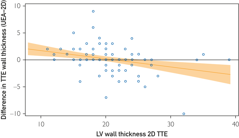
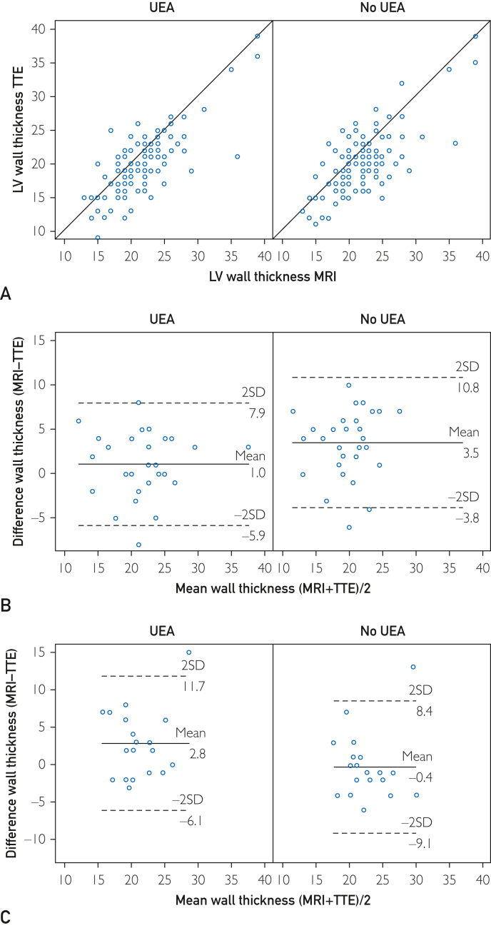
Most (63%) of cardiac imagers found UEA helpful in determining maximal WT by TTE, with 49% reporting change in WT. Of 52 patients where UEA changed WT measurement, 32 (62%) reported an increase and 20 (38%) reported a decrease in WT. The UEA did not alter the median discrepancy in WT between MRI and TTE. However, where UEA increased reported WT, the difference between MRI and TTE improved in 79% of cases (P=.001) from 2.0-0.5mm. In those with scar on MRI, UEA improved agreement of WT between TTE and MRI compared with that of TTE without UEA (79% vs 39%; P=.011). Interclass correlation coefficient for WT for TTE without UEA, with UEA, and MRI was 0.84; (95% CI, 0.61-0.92), 0.88; (95%CI, 0.82-0.92), and 0.97; (95%CI, 0.96-0.98), respectively.
Conclusion
Although use of UEA did not eliminate differences in WT discrepancy between modalities, the addition of UEA to TTE aided in WT determination and improved correlation with MRI in those with greater WT and in all patients with myocardial scars.

 求助内容:
求助内容: 应助结果提醒方式:


