Yahya Efe Guner, Ayhan Comert, Aydın Aslan, Yigit Gungor
{"title":"胼胝体面积和切片:一项与MRI和尸体形态计量学相关的放射解剖学研究。","authors":"Yahya Efe Guner, Ayhan Comert, Aydın Aslan, Yigit Gungor","doi":"10.1007/s00276-023-03206-8","DOIUrl":null,"url":null,"abstract":"<p><strong>Purpose: </strong>The corpus callosum (CC) is the primary interhemispheric connection between the two cerebral hemispheres. Besides their similar morphological characters, there are differences in their measurements. This study aimed to divide the CC into groups using planes based on the anterior commissure (AC) and posterior commissure (PC) and to detect differences in CC magnetic resonance imaging (MRI) and cadaver samples between these groups.</p><p><strong>Methods: </strong>The study included 80 patients (40 male and 40 female patients) who underwent normal MRI in the midsagittal plane, and 38 cerebral hemispheres from 40 adult cadaver brains, with each hemisected in the midsagittal plane. The medial surface of the CC was divided vertically into three parts (the anterior, middle, and posterior zones) according to the AC and PC. Areas and parameters were measured in both the cadaveric hemispheres and patient MRI images.</p><p><strong>Results: </strong>The total CC area and CC areas between, anterior, and posterior to the AC-PC vertical lines were the same in both the MRI and cadaver samples. In addition, morphometric measurements like the CC length, AC-PC length, and CC height at the AC and PC vertical lines, and their correlations were also found to be similar between the MRI and cadaver samples.</p><p><strong>Conclusion: </strong>This study proposes three areas according to AC and PC classification (anterior, middle, and posterior). This new proposed classification is suitable for stereotactic interventions and is useful for obtaining data from MRI images. However, it should be kept in mind that there may be changes and variations.</p>","PeriodicalId":49296,"journal":{"name":"Surgical and Radiologic Anatomy","volume":" ","pages":"1427-1433"},"PeriodicalIF":1.2000,"publicationDate":"2023-11-01","publicationTypes":"Journal Article","fieldsOfStudy":null,"isOpenAccess":false,"openAccessPdf":"","citationCount":"0","resultStr":"{\"title\":\"Corpus callosum area and sectioning: a radioanatomical study correlated with MRI and cadaver morphometry.\",\"authors\":\"Yahya Efe Guner, Ayhan Comert, Aydın Aslan, Yigit Gungor\",\"doi\":\"10.1007/s00276-023-03206-8\",\"DOIUrl\":null,\"url\":null,\"abstract\":\"<p><strong>Purpose: </strong>The corpus callosum (CC) is the primary interhemispheric connection between the two cerebral hemispheres. Besides their similar morphological characters, there are differences in their measurements. This study aimed to divide the CC into groups using planes based on the anterior commissure (AC) and posterior commissure (PC) and to detect differences in CC magnetic resonance imaging (MRI) and cadaver samples between these groups.</p><p><strong>Methods: </strong>The study included 80 patients (40 male and 40 female patients) who underwent normal MRI in the midsagittal plane, and 38 cerebral hemispheres from 40 adult cadaver brains, with each hemisected in the midsagittal plane. The medial surface of the CC was divided vertically into three parts (the anterior, middle, and posterior zones) according to the AC and PC. Areas and parameters were measured in both the cadaveric hemispheres and patient MRI images.</p><p><strong>Results: </strong>The total CC area and CC areas between, anterior, and posterior to the AC-PC vertical lines were the same in both the MRI and cadaver samples. In addition, morphometric measurements like the CC length, AC-PC length, and CC height at the AC and PC vertical lines, and their correlations were also found to be similar between the MRI and cadaver samples.</p><p><strong>Conclusion: </strong>This study proposes three areas according to AC and PC classification (anterior, middle, and posterior). This new proposed classification is suitable for stereotactic interventions and is useful for obtaining data from MRI images. However, it should be kept in mind that there may be changes and variations.</p>\",\"PeriodicalId\":49296,\"journal\":{\"name\":\"Surgical and Radiologic Anatomy\",\"volume\":\" \",\"pages\":\"1427-1433\"},\"PeriodicalIF\":1.2000,\"publicationDate\":\"2023-11-01\",\"publicationTypes\":\"Journal Article\",\"fieldsOfStudy\":null,\"isOpenAccess\":false,\"openAccessPdf\":\"\",\"citationCount\":\"0\",\"resultStr\":null,\"platform\":\"Semanticscholar\",\"paperid\":null,\"PeriodicalName\":\"Surgical and Radiologic Anatomy\",\"FirstCategoryId\":\"3\",\"ListUrlMain\":\"https://doi.org/10.1007/s00276-023-03206-8\",\"RegionNum\":4,\"RegionCategory\":\"医学\",\"ArticlePicture\":[],\"TitleCN\":null,\"AbstractTextCN\":null,\"PMCID\":null,\"EPubDate\":\"2023/7/24 0:00:00\",\"PubModel\":\"Epub\",\"JCR\":\"Q3\",\"JCRName\":\"ANATOMY & MORPHOLOGY\",\"Score\":null,\"Total\":0}","platform":"Semanticscholar","paperid":null,"PeriodicalName":"Surgical and Radiologic Anatomy","FirstCategoryId":"3","ListUrlMain":"https://doi.org/10.1007/s00276-023-03206-8","RegionNum":4,"RegionCategory":"医学","ArticlePicture":[],"TitleCN":null,"AbstractTextCN":null,"PMCID":null,"EPubDate":"2023/7/24 0:00:00","PubModel":"Epub","JCR":"Q3","JCRName":"ANATOMY & MORPHOLOGY","Score":null,"Total":0}
Corpus callosum area and sectioning: a radioanatomical study correlated with MRI and cadaver morphometry.
Purpose: The corpus callosum (CC) is the primary interhemispheric connection between the two cerebral hemispheres. Besides their similar morphological characters, there are differences in their measurements. This study aimed to divide the CC into groups using planes based on the anterior commissure (AC) and posterior commissure (PC) and to detect differences in CC magnetic resonance imaging (MRI) and cadaver samples between these groups.
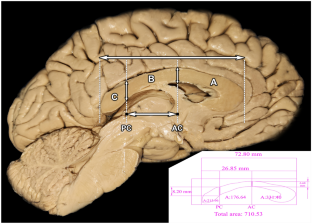
Methods: The study included 80 patients (40 male and 40 female patients) who underwent normal MRI in the midsagittal plane, and 38 cerebral hemispheres from 40 adult cadaver brains, with each hemisected in the midsagittal plane. The medial surface of the CC was divided vertically into three parts (the anterior, middle, and posterior zones) according to the AC and PC. Areas and parameters were measured in both the cadaveric hemispheres and patient MRI images.
Results: The total CC area and CC areas between, anterior, and posterior to the AC-PC vertical lines were the same in both the MRI and cadaver samples. In addition, morphometric measurements like the CC length, AC-PC length, and CC height at the AC and PC vertical lines, and their correlations were also found to be similar between the MRI and cadaver samples.
Conclusion: This study proposes three areas according to AC and PC classification (anterior, middle, and posterior). This new proposed classification is suitable for stereotactic interventions and is useful for obtaining data from MRI images. However, it should be kept in mind that there may be changes and variations.
期刊介绍:
Anatomy is a morphological science which cannot fail to interest the clinician. The practical application of anatomical research to clinical problems necessitates special adaptation and selectivity in choosing from numerous international works. Although there is a tendency to believe that meaningful advances in anatomy are unlikely, constant revision is necessary. Surgical and Radiologic Anatomy, the first international journal of Clinical anatomy has been created in this spirit.
Its goal is to serve clinicians, regardless of speciality-physicians, surgeons, radiologists or other specialists-as an indispensable aid with which they can improve their knowledge of anatomy. Each issue includes: Original papers, review articles, articles on the anatomical bases of medical, surgical and radiological techniques, articles of normal radiologic anatomy, brief reviews of anatomical publications of clinical interest.
Particular attention is given to high quality illustrations, which are indispensable for a better understanding of anatomical problems.
Surgical and Radiologic Anatomy is a journal written by anatomists for clinicians with a special interest in anatomy.

 求助内容:
求助内容: 应助结果提醒方式:


