A Daniel Davidar, Brendan F Judy, Andrew M Hersh, Carly Weber-Levine, Safwan Alomari, Arjun K Menta, Kelly Jiang, Meghana Bhimreddy, Mir Hussain, Neil R Crawford, Majid Khan, Gary Gong, Nicholas Theodore
{"title":"利用基于磁共振成像的合成计算机断层扫描在尸体中进行机器人辅助螺钉固定:朝向无辐射脊柱手术。说明性案例。","authors":"A Daniel Davidar, Brendan F Judy, Andrew M Hersh, Carly Weber-Levine, Safwan Alomari, Arjun K Menta, Kelly Jiang, Meghana Bhimreddy, Mir Hussain, Neil R Crawford, Majid Khan, Gary Gong, Nicholas Theodore","doi":"10.3171/CASE23120","DOIUrl":null,"url":null,"abstract":"<p><strong>Background: </strong>Synthetic computed tomography (sCT) can be created from magnetic resonance imaging (MRI) utilizing newer software. sCT is yet to be explored as a possible alternative to routine CT (rCT). In this study, rCT scans and MRI-derived sCT scans were obtained on a cadaver. Morphometric analysis was performed comparing the 2 scans. The ExcelsiusGPS robot was used to place lumbosacral screws with both rCT and sCT images.</p><p><strong>Observations: </strong>In total, 14 screws were placed. All screws were grade A on the Gertzbein-Robbins scale. The mean surface distance difference between rCT and sCT on a reconstructed software model was -0.02 ± 0.05 mm, the mean absolute surface distance was 0.24 ± 0.05 mm, and the mean absolute error of radiodensity was 92.88 ± 10.53 HU. The overall mean tip distance for the sCT versus rCT was 1.74 ± 1.1 versus 2.36 ± 1.6 mm (p = 0.24); mean tail distance for the sCT versus rCT was 1.93 ± 0.88 versus 2.81 ± 1.03 mm (p = 0.07); and mean angular deviation for the sCT versus rCT was 3.2° ± 2.05° versus 4.04°± 2.71° (p = 0.53).</p><p><strong>Lessons: </strong>MRI-based sCT yielded results comparable to those of rCT in both morphometric analysis and robot-assisted lumbosacral screw placement in a cadaver study.</p>","PeriodicalId":16554,"journal":{"name":"Journal of Neurosurgery: Case Lessons","volume":"6 2","pages":""},"PeriodicalIF":0.0000,"publicationDate":"2023-07-10","publicationTypes":"Journal Article","fieldsOfStudy":null,"isOpenAccess":false,"openAccessPdf":"https://ftp.ncbi.nlm.nih.gov/pub/pmc/oa_pdf/49/c3/CASE23120.PMC10555644.pdf","citationCount":"1","resultStr":"{\"title\":\"Robot-assisted screw fixation in a cadaver utilizing magnetic resonance imaging-based synthetic computed tomography: toward radiation-free spine surgery. Illustrative case.\",\"authors\":\"A Daniel Davidar, Brendan F Judy, Andrew M Hersh, Carly Weber-Levine, Safwan Alomari, Arjun K Menta, Kelly Jiang, Meghana Bhimreddy, Mir Hussain, Neil R Crawford, Majid Khan, Gary Gong, Nicholas Theodore\",\"doi\":\"10.3171/CASE23120\",\"DOIUrl\":null,\"url\":null,\"abstract\":\"<p><strong>Background: </strong>Synthetic computed tomography (sCT) can be created from magnetic resonance imaging (MRI) utilizing newer software. sCT is yet to be explored as a possible alternative to routine CT (rCT). In this study, rCT scans and MRI-derived sCT scans were obtained on a cadaver. Morphometric analysis was performed comparing the 2 scans. The ExcelsiusGPS robot was used to place lumbosacral screws with both rCT and sCT images.</p><p><strong>Observations: </strong>In total, 14 screws were placed. All screws were grade A on the Gertzbein-Robbins scale. The mean surface distance difference between rCT and sCT on a reconstructed software model was -0.02 ± 0.05 mm, the mean absolute surface distance was 0.24 ± 0.05 mm, and the mean absolute error of radiodensity was 92.88 ± 10.53 HU. The overall mean tip distance for the sCT versus rCT was 1.74 ± 1.1 versus 2.36 ± 1.6 mm (p = 0.24); mean tail distance for the sCT versus rCT was 1.93 ± 0.88 versus 2.81 ± 1.03 mm (p = 0.07); and mean angular deviation for the sCT versus rCT was 3.2° ± 2.05° versus 4.04°± 2.71° (p = 0.53).</p><p><strong>Lessons: </strong>MRI-based sCT yielded results comparable to those of rCT in both morphometric analysis and robot-assisted lumbosacral screw placement in a cadaver study.</p>\",\"PeriodicalId\":16554,\"journal\":{\"name\":\"Journal of Neurosurgery: Case Lessons\",\"volume\":\"6 2\",\"pages\":\"\"},\"PeriodicalIF\":0.0000,\"publicationDate\":\"2023-07-10\",\"publicationTypes\":\"Journal Article\",\"fieldsOfStudy\":null,\"isOpenAccess\":false,\"openAccessPdf\":\"https://ftp.ncbi.nlm.nih.gov/pub/pmc/oa_pdf/49/c3/CASE23120.PMC10555644.pdf\",\"citationCount\":\"1\",\"resultStr\":null,\"platform\":\"Semanticscholar\",\"paperid\":null,\"PeriodicalName\":\"Journal of Neurosurgery: Case Lessons\",\"FirstCategoryId\":\"1085\",\"ListUrlMain\":\"https://doi.org/10.3171/CASE23120\",\"RegionNum\":0,\"RegionCategory\":null,\"ArticlePicture\":[],\"TitleCN\":null,\"AbstractTextCN\":null,\"PMCID\":null,\"EPubDate\":\"\",\"PubModel\":\"\",\"JCR\":\"\",\"JCRName\":\"\",\"Score\":null,\"Total\":0}","platform":"Semanticscholar","paperid":null,"PeriodicalName":"Journal of Neurosurgery: Case Lessons","FirstCategoryId":"1085","ListUrlMain":"https://doi.org/10.3171/CASE23120","RegionNum":0,"RegionCategory":null,"ArticlePicture":[],"TitleCN":null,"AbstractTextCN":null,"PMCID":null,"EPubDate":"","PubModel":"","JCR":"","JCRName":"","Score":null,"Total":0}
Robot-assisted screw fixation in a cadaver utilizing magnetic resonance imaging-based synthetic computed tomography: toward radiation-free spine surgery. Illustrative case.
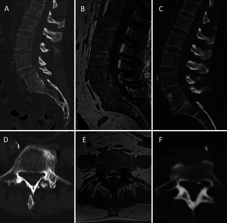
Background: Synthetic computed tomography (sCT) can be created from magnetic resonance imaging (MRI) utilizing newer software. sCT is yet to be explored as a possible alternative to routine CT (rCT). In this study, rCT scans and MRI-derived sCT scans were obtained on a cadaver. Morphometric analysis was performed comparing the 2 scans. The ExcelsiusGPS robot was used to place lumbosacral screws with both rCT and sCT images.
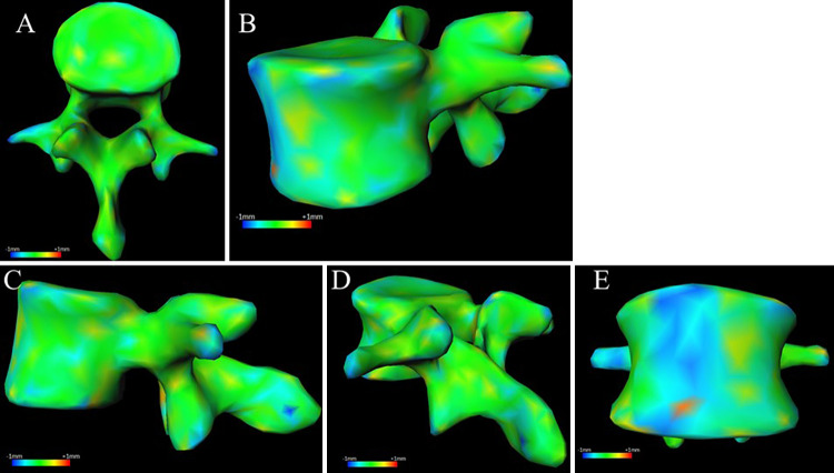
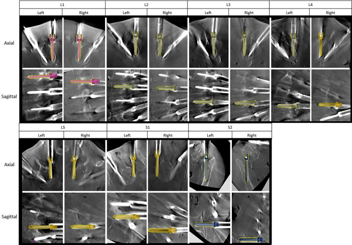
Observations: In total, 14 screws were placed. All screws were grade A on the Gertzbein-Robbins scale. The mean surface distance difference between rCT and sCT on a reconstructed software model was -0.02 ± 0.05 mm, the mean absolute surface distance was 0.24 ± 0.05 mm, and the mean absolute error of radiodensity was 92.88 ± 10.53 HU. The overall mean tip distance for the sCT versus rCT was 1.74 ± 1.1 versus 2.36 ± 1.6 mm (p = 0.24); mean tail distance for the sCT versus rCT was 1.93 ± 0.88 versus 2.81 ± 1.03 mm (p = 0.07); and mean angular deviation for the sCT versus rCT was 3.2° ± 2.05° versus 4.04°± 2.71° (p = 0.53).
Lessons: MRI-based sCT yielded results comparable to those of rCT in both morphometric analysis and robot-assisted lumbosacral screw placement in a cadaver study.

 求助内容:
求助内容: 应助结果提醒方式:


