{"title":"肝外胆管癌神经周围浸润的三维组织清除分析。","authors":"Hirokazu Ogasawara, Tadashi Yoshizawa, Kiyoko Oshima, Kenta Ogasawara, Shunsuke Kubota, Shintaro Goto, Satoko Morohashi, Taiichi Wakiya, Norihisa Kimura, Keinosuke Ishido, Hiroshi Kijima, Kenichi Hakamada","doi":"10.3389/pore.2023.1611284","DOIUrl":null,"url":null,"abstract":"<p><p>Perineural invasion (PNI) is a characteristic invasion pattern of distal cholangiocarcinoma (DCC). Conventional histopathologic examination is a challenging approach to analyze the spatial relationship between cancer and neural tissue in full-thickness bile duct specimens. Therefore, we used a tissue clearing method to examine PNI in DCC with three-dimensional (3D) structural analysis. The immunolabeling-enabled 3D imaging of solvent-cleared organs method was performed to examine 20 DCC specimens from five patients and 8 non-neoplastic bile duct specimens from two controls. The bile duct epithelium and neural tissue were labeled with CK19 and S100 antibodies, respectively. Two-dimensional hematoxylin/eosin staining revealed only PNI around thick nerve fibers in the deep layer of the bile duct, whereas PNI was not identified in the superficial layer. 3D analysis revealed that the parts of DCC closer to the mucosa exhibited more nerves than the normal bile duct. The nerve fibers were continuously branched and connected with thick nerve fibers in the deep layer of the bile duct. DCC formed a tubular structure invading from the epithelium and extending around thin nerve fibers in the superficial layer. DCC exhibited continuous infiltration around the thick nerve fibers in the deep layer. This is the first study using a tissue clearing method to examine the PNI of DCC, providing new insights into the underlying mechanisms.</p>","PeriodicalId":19981,"journal":{"name":"Pathology & Oncology Research","volume":"29 ","pages":"1611284"},"PeriodicalIF":2.3000,"publicationDate":"2023-01-01","publicationTypes":"Journal Article","fieldsOfStudy":null,"isOpenAccess":false,"openAccessPdf":"https://www.ncbi.nlm.nih.gov/pmc/articles/PMC10323134/pdf/","citationCount":"0","resultStr":"{\"title\":\"Three-dimensional analysis of perineural invasion in extrahepatic cholangiocarcinoma using tissue clearing.\",\"authors\":\"Hirokazu Ogasawara, Tadashi Yoshizawa, Kiyoko Oshima, Kenta Ogasawara, Shunsuke Kubota, Shintaro Goto, Satoko Morohashi, Taiichi Wakiya, Norihisa Kimura, Keinosuke Ishido, Hiroshi Kijima, Kenichi Hakamada\",\"doi\":\"10.3389/pore.2023.1611284\",\"DOIUrl\":null,\"url\":null,\"abstract\":\"<p><p>Perineural invasion (PNI) is a characteristic invasion pattern of distal cholangiocarcinoma (DCC). Conventional histopathologic examination is a challenging approach to analyze the spatial relationship between cancer and neural tissue in full-thickness bile duct specimens. Therefore, we used a tissue clearing method to examine PNI in DCC with three-dimensional (3D) structural analysis. The immunolabeling-enabled 3D imaging of solvent-cleared organs method was performed to examine 20 DCC specimens from five patients and 8 non-neoplastic bile duct specimens from two controls. The bile duct epithelium and neural tissue were labeled with CK19 and S100 antibodies, respectively. Two-dimensional hematoxylin/eosin staining revealed only PNI around thick nerve fibers in the deep layer of the bile duct, whereas PNI was not identified in the superficial layer. 3D analysis revealed that the parts of DCC closer to the mucosa exhibited more nerves than the normal bile duct. The nerve fibers were continuously branched and connected with thick nerve fibers in the deep layer of the bile duct. DCC formed a tubular structure invading from the epithelium and extending around thin nerve fibers in the superficial layer. DCC exhibited continuous infiltration around the thick nerve fibers in the deep layer. This is the first study using a tissue clearing method to examine the PNI of DCC, providing new insights into the underlying mechanisms.</p>\",\"PeriodicalId\":19981,\"journal\":{\"name\":\"Pathology & Oncology Research\",\"volume\":\"29 \",\"pages\":\"1611284\"},\"PeriodicalIF\":2.3000,\"publicationDate\":\"2023-01-01\",\"publicationTypes\":\"Journal Article\",\"fieldsOfStudy\":null,\"isOpenAccess\":false,\"openAccessPdf\":\"https://www.ncbi.nlm.nih.gov/pmc/articles/PMC10323134/pdf/\",\"citationCount\":\"0\",\"resultStr\":null,\"platform\":\"Semanticscholar\",\"paperid\":null,\"PeriodicalName\":\"Pathology & Oncology Research\",\"FirstCategoryId\":\"3\",\"ListUrlMain\":\"https://doi.org/10.3389/pore.2023.1611284\",\"RegionNum\":4,\"RegionCategory\":\"医学\",\"ArticlePicture\":[],\"TitleCN\":null,\"AbstractTextCN\":null,\"PMCID\":null,\"EPubDate\":\"\",\"PubModel\":\"\",\"JCR\":\"Q3\",\"JCRName\":\"ONCOLOGY\",\"Score\":null,\"Total\":0}","platform":"Semanticscholar","paperid":null,"PeriodicalName":"Pathology & Oncology Research","FirstCategoryId":"3","ListUrlMain":"https://doi.org/10.3389/pore.2023.1611284","RegionNum":4,"RegionCategory":"医学","ArticlePicture":[],"TitleCN":null,"AbstractTextCN":null,"PMCID":null,"EPubDate":"","PubModel":"","JCR":"Q3","JCRName":"ONCOLOGY","Score":null,"Total":0}
Three-dimensional analysis of perineural invasion in extrahepatic cholangiocarcinoma using tissue clearing.
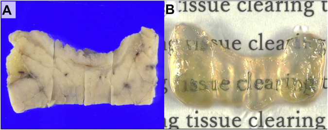
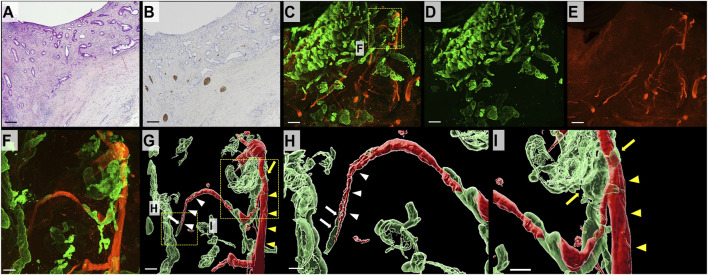
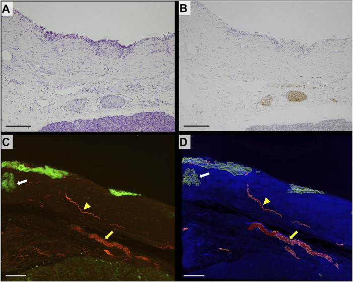
Perineural invasion (PNI) is a characteristic invasion pattern of distal cholangiocarcinoma (DCC). Conventional histopathologic examination is a challenging approach to analyze the spatial relationship between cancer and neural tissue in full-thickness bile duct specimens. Therefore, we used a tissue clearing method to examine PNI in DCC with three-dimensional (3D) structural analysis. The immunolabeling-enabled 3D imaging of solvent-cleared organs method was performed to examine 20 DCC specimens from five patients and 8 non-neoplastic bile duct specimens from two controls. The bile duct epithelium and neural tissue were labeled with CK19 and S100 antibodies, respectively. Two-dimensional hematoxylin/eosin staining revealed only PNI around thick nerve fibers in the deep layer of the bile duct, whereas PNI was not identified in the superficial layer. 3D analysis revealed that the parts of DCC closer to the mucosa exhibited more nerves than the normal bile duct. The nerve fibers were continuously branched and connected with thick nerve fibers in the deep layer of the bile duct. DCC formed a tubular structure invading from the epithelium and extending around thin nerve fibers in the superficial layer. DCC exhibited continuous infiltration around the thick nerve fibers in the deep layer. This is the first study using a tissue clearing method to examine the PNI of DCC, providing new insights into the underlying mechanisms.
期刊介绍:
Pathology & Oncology Research (POR) is an interdisciplinary Journal at the interface of pathology and oncology including the preclinical and translational research, diagnostics and therapy. Furthermore, POR is an international forum for the rapid communication of reviews, original research, critical and topical reports with excellence and novelty. Published quarterly, POR is dedicated to keeping scientists informed of developments on the selected biomedical fields bridging the gap between basic research and clinical medicine. It is a special aim for POR to promote pathological and oncological publishing activity of colleagues in the Central and East European region. The journal will be of interest to pathologists, and a broad range of experimental and clinical oncologists, and related experts. POR is supported by an acknowledged international advisory board and the Arányi Fundation for modern pathology.
 求助内容:
求助内容: 应助结果提醒方式:


