Katherine Callahan, Isidora Beach, Sadie Casale, John DeWitt, Bruce Tranmer
{"title":"肿瘤内脓肿合并放疗后世界卫生组织二级脑膜瘤:一个例证性病例。","authors":"Katherine Callahan, Isidora Beach, Sadie Casale, John DeWitt, Bruce Tranmer","doi":"10.3171/CASE23146","DOIUrl":null,"url":null,"abstract":"<p><strong>Background: </strong>Cerebral meningiomas and brain abscesses are common independently, but intrameningioma abscesses rarely occur, with only 15 cases in the literature. These abscesses most frequently develop in patients with a known source of bacteremia; only one case of intrameningioma abscess without a known source of infection has been reported previously.</p><p><strong>Observations: </strong>This is the second reported case of an intrameningioma abscess without a clear source of infection, occurring in a 70-year-old female with a history of transsphenoidal craniopharyngioma resection and radiation many years prior. She presented with severe fatigue and altered mental status initially ascribed to adrenal insufficiency, and magnetic resonance imaging showed a new heterogeneously enhancing left temporal mass with surrounding edema. After urgent tumor resection, pathology demonstrated a World Health Organization grade II meningioma (radiation induced). After a course of steroids and intravenous nafcillin, the patient recovered without neurological deficits.</p><p><strong>Lessons: </strong>The natural history of intrameningioma abscesses is not fully understood. These uncommon lesions can form secondary to hematogenous spread facilitated by meningiomas' robust vascularization, typically in patients with bacteremia. Even when no significant source of infection is identified, the differential diagnosis of intrameningioma abscess should be considered because this pathology can be rapidly progressive, even fatal, but is treatable if recognized promptly.</p>","PeriodicalId":16554,"journal":{"name":"Journal of Neurosurgery: Case Lessons","volume":"6 1","pages":""},"PeriodicalIF":0.0000,"publicationDate":"2023-07-03","publicationTypes":"Journal Article","fieldsOfStudy":null,"isOpenAccess":false,"openAccessPdf":"https://ftp.ncbi.nlm.nih.gov/pub/pmc/oa_pdf/53/ea/CASE23146.PMC10555632.pdf","citationCount":"0","resultStr":"{\"title\":\"Intratumoral abscess complicating a postradiation-induced World Health Organization grade II meningioma: illustrative case.\",\"authors\":\"Katherine Callahan, Isidora Beach, Sadie Casale, John DeWitt, Bruce Tranmer\",\"doi\":\"10.3171/CASE23146\",\"DOIUrl\":null,\"url\":null,\"abstract\":\"<p><strong>Background: </strong>Cerebral meningiomas and brain abscesses are common independently, but intrameningioma abscesses rarely occur, with only 15 cases in the literature. These abscesses most frequently develop in patients with a known source of bacteremia; only one case of intrameningioma abscess without a known source of infection has been reported previously.</p><p><strong>Observations: </strong>This is the second reported case of an intrameningioma abscess without a clear source of infection, occurring in a 70-year-old female with a history of transsphenoidal craniopharyngioma resection and radiation many years prior. She presented with severe fatigue and altered mental status initially ascribed to adrenal insufficiency, and magnetic resonance imaging showed a new heterogeneously enhancing left temporal mass with surrounding edema. After urgent tumor resection, pathology demonstrated a World Health Organization grade II meningioma (radiation induced). After a course of steroids and intravenous nafcillin, the patient recovered without neurological deficits.</p><p><strong>Lessons: </strong>The natural history of intrameningioma abscesses is not fully understood. These uncommon lesions can form secondary to hematogenous spread facilitated by meningiomas' robust vascularization, typically in patients with bacteremia. Even when no significant source of infection is identified, the differential diagnosis of intrameningioma abscess should be considered because this pathology can be rapidly progressive, even fatal, but is treatable if recognized promptly.</p>\",\"PeriodicalId\":16554,\"journal\":{\"name\":\"Journal of Neurosurgery: Case Lessons\",\"volume\":\"6 1\",\"pages\":\"\"},\"PeriodicalIF\":0.0000,\"publicationDate\":\"2023-07-03\",\"publicationTypes\":\"Journal Article\",\"fieldsOfStudy\":null,\"isOpenAccess\":false,\"openAccessPdf\":\"https://ftp.ncbi.nlm.nih.gov/pub/pmc/oa_pdf/53/ea/CASE23146.PMC10555632.pdf\",\"citationCount\":\"0\",\"resultStr\":null,\"platform\":\"Semanticscholar\",\"paperid\":null,\"PeriodicalName\":\"Journal of Neurosurgery: Case Lessons\",\"FirstCategoryId\":\"1085\",\"ListUrlMain\":\"https://doi.org/10.3171/CASE23146\",\"RegionNum\":0,\"RegionCategory\":null,\"ArticlePicture\":[],\"TitleCN\":null,\"AbstractTextCN\":null,\"PMCID\":null,\"EPubDate\":\"\",\"PubModel\":\"\",\"JCR\":\"\",\"JCRName\":\"\",\"Score\":null,\"Total\":0}","platform":"Semanticscholar","paperid":null,"PeriodicalName":"Journal of Neurosurgery: Case Lessons","FirstCategoryId":"1085","ListUrlMain":"https://doi.org/10.3171/CASE23146","RegionNum":0,"RegionCategory":null,"ArticlePicture":[],"TitleCN":null,"AbstractTextCN":null,"PMCID":null,"EPubDate":"","PubModel":"","JCR":"","JCRName":"","Score":null,"Total":0}
Intratumoral abscess complicating a postradiation-induced World Health Organization grade II meningioma: illustrative case.
Background: Cerebral meningiomas and brain abscesses are common independently, but intrameningioma abscesses rarely occur, with only 15 cases in the literature. These abscesses most frequently develop in patients with a known source of bacteremia; only one case of intrameningioma abscess without a known source of infection has been reported previously.
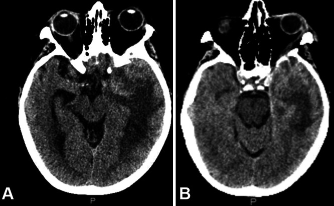
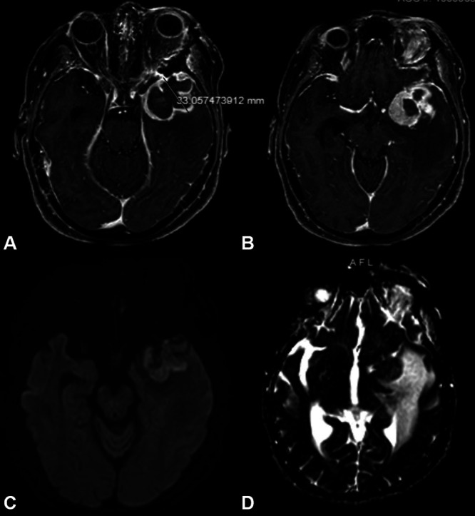
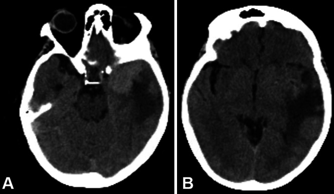
Observations: This is the second reported case of an intrameningioma abscess without a clear source of infection, occurring in a 70-year-old female with a history of transsphenoidal craniopharyngioma resection and radiation many years prior. She presented with severe fatigue and altered mental status initially ascribed to adrenal insufficiency, and magnetic resonance imaging showed a new heterogeneously enhancing left temporal mass with surrounding edema. After urgent tumor resection, pathology demonstrated a World Health Organization grade II meningioma (radiation induced). After a course of steroids and intravenous nafcillin, the patient recovered without neurological deficits.
Lessons: The natural history of intrameningioma abscesses is not fully understood. These uncommon lesions can form secondary to hematogenous spread facilitated by meningiomas' robust vascularization, typically in patients with bacteremia. Even when no significant source of infection is identified, the differential diagnosis of intrameningioma abscess should be considered because this pathology can be rapidly progressive, even fatal, but is treatable if recognized promptly.

 求助内容:
求助内容: 应助结果提醒方式:


