Diogo Tiago da Silva, Maria Luana Alves, Júlio Cesar Pereira Spada, João Augusto Franco Leonel, Geovanna Vioti, Julia Cristina Benassi, Valéria Maria Lara Carregaro, Maria Fernanda Alves-Martin, Wilma Aparecida Starke-Buzetti, Trícia Maria Ferreira de Sousa Oliveira
{"title":"猫利什曼病:血液学和生化分析。","authors":"Diogo Tiago da Silva, Maria Luana Alves, Júlio Cesar Pereira Spada, João Augusto Franco Leonel, Geovanna Vioti, Julia Cristina Benassi, Valéria Maria Lara Carregaro, Maria Fernanda Alves-Martin, Wilma Aparecida Starke-Buzetti, Trícia Maria Ferreira de Sousa Oliveira","doi":"10.1590/S1984-29612023035","DOIUrl":null,"url":null,"abstract":"<p><p>One hundred and sixty-six cats from two animal shelters were subjected to enzyme-linked immunosorbent assay (ELISA), indirect immunofluorescence antibody test (IFAT), conventional polymerase chain reaction (cPCR), quantitative PCR (qPCR) and parasitological tests (PA) for the diagnosis of Leishmania spp. Among them, 15% (25/166), 53.6% (89/166), 3.6% (06/166) and 1.8% (03/166) were positive by ELISA, IFAT, both PCRs and PA, respectively. The sequencing of ITS-1 PCR amplicons revealed a 100% match with Leishmania infantum. After the Leishmania spp. survey, 12 cats were selected and divided into two groups for clinical, hematological, and biochemical analysis: six L. infantum positive cats (G1) and six Leishmania spp. negative cats (G2). All the cats were negative for feline immunodeficiency virus (FIV) and feline leukemia virus (FeLV). A statistical analysis indicated significantly low platelet counts and significant hyperproteinemia associated with hypoalbuminemia in positive cats (p<0.05). Our results suggest that in endemic areas, cats with clinical signs of feline leishmaniosis (such as skin lesions, weight loss and/or enlarged lymph nodes) and that exhibit hematological and biochemical changes, such as low platelet counts and hyperproteinemia with hypoalbuminemia, should be tested for Leishmania spp. infection.</p>","PeriodicalId":21270,"journal":{"name":"Revista brasileira de parasitologia veterinaria = Brazilian journal of veterinary parasitology : Orgao Oficial do Colegio Brasileiro de Parasitologia Veterinaria","volume":"32 2","pages":"e003823"},"PeriodicalIF":0.0000,"publicationDate":"2023-01-01","publicationTypes":"Journal Article","fieldsOfStudy":null,"isOpenAccess":false,"openAccessPdf":"https://www.ncbi.nlm.nih.gov/pmc/articles/PMC10321720/pdf/","citationCount":"0","resultStr":"{\"title\":\"Feline leishmaniosis: hematological and biochemical analysis.\",\"authors\":\"Diogo Tiago da Silva, Maria Luana Alves, Júlio Cesar Pereira Spada, João Augusto Franco Leonel, Geovanna Vioti, Julia Cristina Benassi, Valéria Maria Lara Carregaro, Maria Fernanda Alves-Martin, Wilma Aparecida Starke-Buzetti, Trícia Maria Ferreira de Sousa Oliveira\",\"doi\":\"10.1590/S1984-29612023035\",\"DOIUrl\":null,\"url\":null,\"abstract\":\"<p><p>One hundred and sixty-six cats from two animal shelters were subjected to enzyme-linked immunosorbent assay (ELISA), indirect immunofluorescence antibody test (IFAT), conventional polymerase chain reaction (cPCR), quantitative PCR (qPCR) and parasitological tests (PA) for the diagnosis of Leishmania spp. Among them, 15% (25/166), 53.6% (89/166), 3.6% (06/166) and 1.8% (03/166) were positive by ELISA, IFAT, both PCRs and PA, respectively. The sequencing of ITS-1 PCR amplicons revealed a 100% match with Leishmania infantum. After the Leishmania spp. survey, 12 cats were selected and divided into two groups for clinical, hematological, and biochemical analysis: six L. infantum positive cats (G1) and six Leishmania spp. negative cats (G2). All the cats were negative for feline immunodeficiency virus (FIV) and feline leukemia virus (FeLV). A statistical analysis indicated significantly low platelet counts and significant hyperproteinemia associated with hypoalbuminemia in positive cats (p<0.05). Our results suggest that in endemic areas, cats with clinical signs of feline leishmaniosis (such as skin lesions, weight loss and/or enlarged lymph nodes) and that exhibit hematological and biochemical changes, such as low platelet counts and hyperproteinemia with hypoalbuminemia, should be tested for Leishmania spp. infection.</p>\",\"PeriodicalId\":21270,\"journal\":{\"name\":\"Revista brasileira de parasitologia veterinaria = Brazilian journal of veterinary parasitology : Orgao Oficial do Colegio Brasileiro de Parasitologia Veterinaria\",\"volume\":\"32 2\",\"pages\":\"e003823\"},\"PeriodicalIF\":0.0000,\"publicationDate\":\"2023-01-01\",\"publicationTypes\":\"Journal Article\",\"fieldsOfStudy\":null,\"isOpenAccess\":false,\"openAccessPdf\":\"https://www.ncbi.nlm.nih.gov/pmc/articles/PMC10321720/pdf/\",\"citationCount\":\"0\",\"resultStr\":null,\"platform\":\"Semanticscholar\",\"paperid\":null,\"PeriodicalName\":\"Revista brasileira de parasitologia veterinaria = Brazilian journal of veterinary parasitology : Orgao Oficial do Colegio Brasileiro de Parasitologia Veterinaria\",\"FirstCategoryId\":\"1085\",\"ListUrlMain\":\"https://doi.org/10.1590/S1984-29612023035\",\"RegionNum\":0,\"RegionCategory\":null,\"ArticlePicture\":[],\"TitleCN\":null,\"AbstractTextCN\":null,\"PMCID\":null,\"EPubDate\":\"\",\"PubModel\":\"\",\"JCR\":\"\",\"JCRName\":\"\",\"Score\":null,\"Total\":0}","platform":"Semanticscholar","paperid":null,"PeriodicalName":"Revista brasileira de parasitologia veterinaria = Brazilian journal of veterinary parasitology : Orgao Oficial do Colegio Brasileiro de Parasitologia Veterinaria","FirstCategoryId":"1085","ListUrlMain":"https://doi.org/10.1590/S1984-29612023035","RegionNum":0,"RegionCategory":null,"ArticlePicture":[],"TitleCN":null,"AbstractTextCN":null,"PMCID":null,"EPubDate":"","PubModel":"","JCR":"","JCRName":"","Score":null,"Total":0}
Feline leishmaniosis: hematological and biochemical analysis.
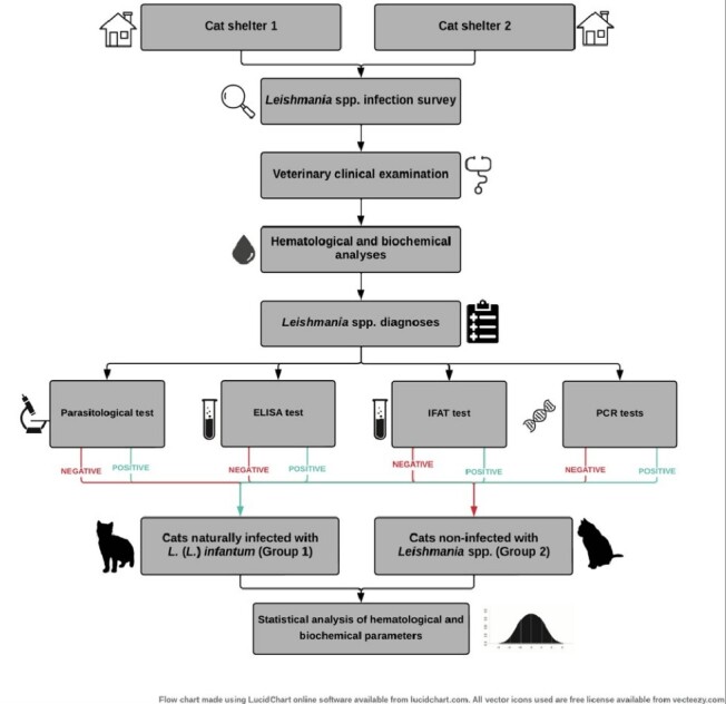
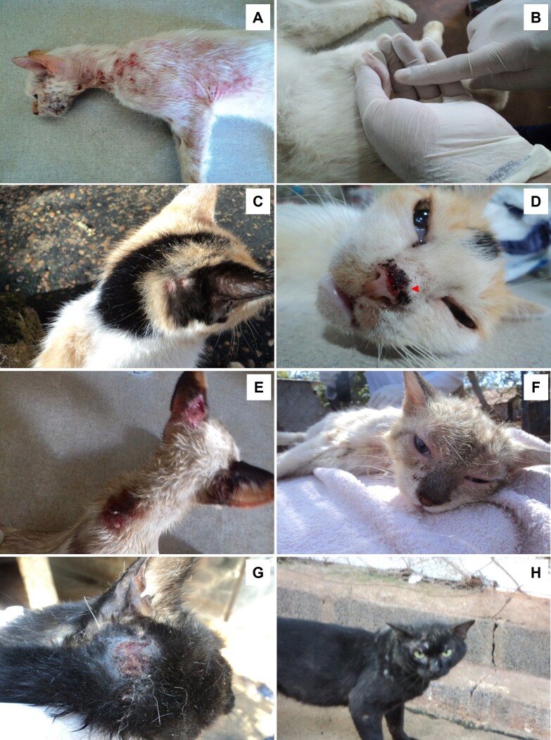
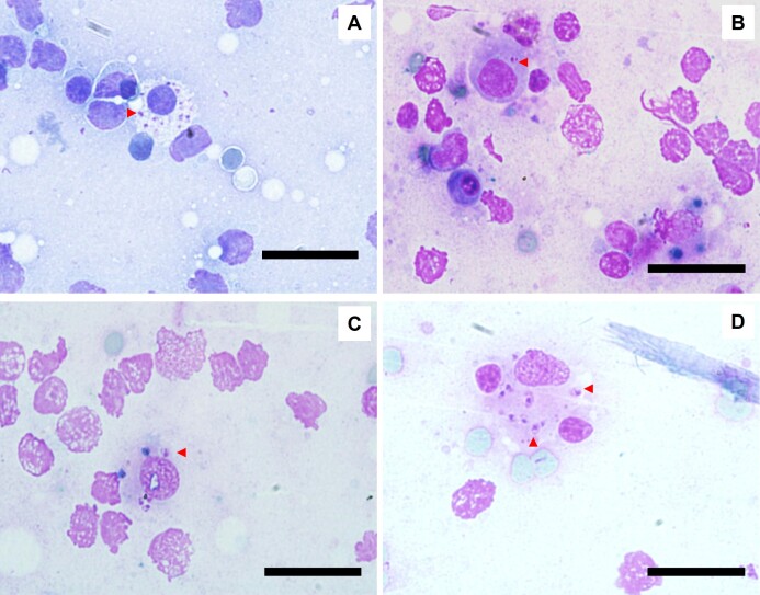
One hundred and sixty-six cats from two animal shelters were subjected to enzyme-linked immunosorbent assay (ELISA), indirect immunofluorescence antibody test (IFAT), conventional polymerase chain reaction (cPCR), quantitative PCR (qPCR) and parasitological tests (PA) for the diagnosis of Leishmania spp. Among them, 15% (25/166), 53.6% (89/166), 3.6% (06/166) and 1.8% (03/166) were positive by ELISA, IFAT, both PCRs and PA, respectively. The sequencing of ITS-1 PCR amplicons revealed a 100% match with Leishmania infantum. After the Leishmania spp. survey, 12 cats were selected and divided into two groups for clinical, hematological, and biochemical analysis: six L. infantum positive cats (G1) and six Leishmania spp. negative cats (G2). All the cats were negative for feline immunodeficiency virus (FIV) and feline leukemia virus (FeLV). A statistical analysis indicated significantly low platelet counts and significant hyperproteinemia associated with hypoalbuminemia in positive cats (p<0.05). Our results suggest that in endemic areas, cats with clinical signs of feline leishmaniosis (such as skin lesions, weight loss and/or enlarged lymph nodes) and that exhibit hematological and biochemical changes, such as low platelet counts and hyperproteinemia with hypoalbuminemia, should be tested for Leishmania spp. infection.

 求助内容:
求助内容: 应助结果提醒方式:


