Osvaldo Zmener, Cornelis H Pameijer, Ana C Boetto, Mariana Picca
{"title":"下颌第三磨牙牙釉质的凹陷和裂缝深度:微渗漏的一个开放的大门?","authors":"Osvaldo Zmener, Cornelis H Pameijer, Ana C Boetto, Mariana Picca","doi":"10.54589/aol.34/2/183","DOIUrl":null,"url":null,"abstract":"<p><p>The aim of this descriptive ex vivo study was to evaluate qualitatively the depth of pit and fissures (P&F) of the enamel in human mandibular third molars. Fifty (n=50) extracted human mandibular third molars were cleaned and disinfected. All tooth surfaces were coated with nail varnish except for a 1-mm margin around the periphery of the occlusal surface. The teeth were immersed for 48 hours at 37 °C in 1% methylene blue dye solution prepared in artificial saliva. After cleaning, the crowns were separated from the root at the cementoenamel junction and subsequently sectioned longitudinally in buccolingual direction at the location of the central fossa. All sections were examined using a stereoscopic microscope and photographed. The images were downloaded on a computer. The length of penetration of the P&F was recorded using the following scoring system: C1: P&F extended to half of the enamel thickness; C2: P&F extended beyond half of the enamel thickness without reaching the dentine-enamel junction; C3: P&F extended to the dentine-enamel junction. For pits, C1, C2 and C3 were observed in 35, 9 and 6 teeth, respectively, while for fissures, C1, C2 and C3 were observed in 15, 18 and 17 teeth, respectively. The P&F detected in the samples extended to the deepest portions of enamel, quite frequently reaching the enamel-dentine junction. Clinicians should recognize that even if pits and fissures are not clinically obvious, they penetrate deep into the enamel and frequently reach the dentine-enamel junction. Effective treatment is recommended to block access to P&F, thus preventing ingress of bacteria.</p>","PeriodicalId":7033,"journal":{"name":"Acta odontologica latinoamericana : AOL","volume":"34 2","pages":"183-187"},"PeriodicalIF":0.0000,"publicationDate":"2021-08-01","publicationTypes":"Journal Article","fieldsOfStudy":null,"isOpenAccess":false,"openAccessPdf":"https://ftp.ncbi.nlm.nih.gov/pub/pmc/oa_pdf/f1/eb/1852-4834-34-2-183.PMC10315076.pdf","citationCount":"0","resultStr":"{\"title\":\"Pit and fissure depth in the enamel of mandibular third molars: An open gate for microleakage?\",\"authors\":\"Osvaldo Zmener, Cornelis H Pameijer, Ana C Boetto, Mariana Picca\",\"doi\":\"10.54589/aol.34/2/183\",\"DOIUrl\":null,\"url\":null,\"abstract\":\"<p><p>The aim of this descriptive ex vivo study was to evaluate qualitatively the depth of pit and fissures (P&F) of the enamel in human mandibular third molars. Fifty (n=50) extracted human mandibular third molars were cleaned and disinfected. All tooth surfaces were coated with nail varnish except for a 1-mm margin around the periphery of the occlusal surface. The teeth were immersed for 48 hours at 37 °C in 1% methylene blue dye solution prepared in artificial saliva. After cleaning, the crowns were separated from the root at the cementoenamel junction and subsequently sectioned longitudinally in buccolingual direction at the location of the central fossa. All sections were examined using a stereoscopic microscope and photographed. The images were downloaded on a computer. The length of penetration of the P&F was recorded using the following scoring system: C1: P&F extended to half of the enamel thickness; C2: P&F extended beyond half of the enamel thickness without reaching the dentine-enamel junction; C3: P&F extended to the dentine-enamel junction. For pits, C1, C2 and C3 were observed in 35, 9 and 6 teeth, respectively, while for fissures, C1, C2 and C3 were observed in 15, 18 and 17 teeth, respectively. The P&F detected in the samples extended to the deepest portions of enamel, quite frequently reaching the enamel-dentine junction. Clinicians should recognize that even if pits and fissures are not clinically obvious, they penetrate deep into the enamel and frequently reach the dentine-enamel junction. Effective treatment is recommended to block access to P&F, thus preventing ingress of bacteria.</p>\",\"PeriodicalId\":7033,\"journal\":{\"name\":\"Acta odontologica latinoamericana : AOL\",\"volume\":\"34 2\",\"pages\":\"183-187\"},\"PeriodicalIF\":0.0000,\"publicationDate\":\"2021-08-01\",\"publicationTypes\":\"Journal Article\",\"fieldsOfStudy\":null,\"isOpenAccess\":false,\"openAccessPdf\":\"https://ftp.ncbi.nlm.nih.gov/pub/pmc/oa_pdf/f1/eb/1852-4834-34-2-183.PMC10315076.pdf\",\"citationCount\":\"0\",\"resultStr\":null,\"platform\":\"Semanticscholar\",\"paperid\":null,\"PeriodicalName\":\"Acta odontologica latinoamericana : AOL\",\"FirstCategoryId\":\"1085\",\"ListUrlMain\":\"https://doi.org/10.54589/aol.34/2/183\",\"RegionNum\":0,\"RegionCategory\":null,\"ArticlePicture\":[],\"TitleCN\":null,\"AbstractTextCN\":null,\"PMCID\":null,\"EPubDate\":\"\",\"PubModel\":\"\",\"JCR\":\"\",\"JCRName\":\"\",\"Score\":null,\"Total\":0}","platform":"Semanticscholar","paperid":null,"PeriodicalName":"Acta odontologica latinoamericana : AOL","FirstCategoryId":"1085","ListUrlMain":"https://doi.org/10.54589/aol.34/2/183","RegionNum":0,"RegionCategory":null,"ArticlePicture":[],"TitleCN":null,"AbstractTextCN":null,"PMCID":null,"EPubDate":"","PubModel":"","JCR":"","JCRName":"","Score":null,"Total":0}
Pit and fissure depth in the enamel of mandibular third molars: An open gate for microleakage?
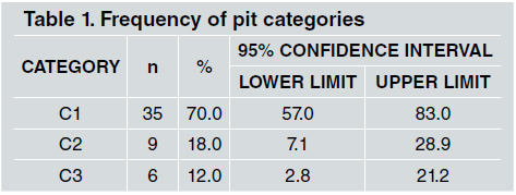
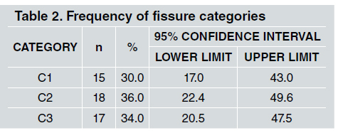
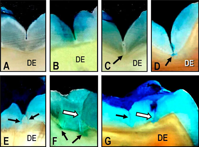
The aim of this descriptive ex vivo study was to evaluate qualitatively the depth of pit and fissures (P&F) of the enamel in human mandibular third molars. Fifty (n=50) extracted human mandibular third molars were cleaned and disinfected. All tooth surfaces were coated with nail varnish except for a 1-mm margin around the periphery of the occlusal surface. The teeth were immersed for 48 hours at 37 °C in 1% methylene blue dye solution prepared in artificial saliva. After cleaning, the crowns were separated from the root at the cementoenamel junction and subsequently sectioned longitudinally in buccolingual direction at the location of the central fossa. All sections were examined using a stereoscopic microscope and photographed. The images were downloaded on a computer. The length of penetration of the P&F was recorded using the following scoring system: C1: P&F extended to half of the enamel thickness; C2: P&F extended beyond half of the enamel thickness without reaching the dentine-enamel junction; C3: P&F extended to the dentine-enamel junction. For pits, C1, C2 and C3 were observed in 35, 9 and 6 teeth, respectively, while for fissures, C1, C2 and C3 were observed in 15, 18 and 17 teeth, respectively. The P&F detected in the samples extended to the deepest portions of enamel, quite frequently reaching the enamel-dentine junction. Clinicians should recognize that even if pits and fissures are not clinically obvious, they penetrate deep into the enamel and frequently reach the dentine-enamel junction. Effective treatment is recommended to block access to P&F, thus preventing ingress of bacteria.

 求助内容:
求助内容: 应助结果提醒方式:


