Ana Indira Bezerra Barros Gadelha, Moacir Franco de Oliveira, Ana Caroline Freitas Caetano de Sousa, João Augusto Rodrigues Alves Diniz, Igor Renno Guimarães Lopes, Bruno Caio Chaves Fernandes, Alexsandra Fernandes Pereira, Carlos Eduardo Bezerra de Moura
{"title":"美洲大黄的胚胎外膜形态(Rhea americana americana Linnaeus,1758)。","authors":"Ana Indira Bezerra Barros Gadelha, Moacir Franco de Oliveira, Ana Caroline Freitas Caetano de Sousa, João Augusto Rodrigues Alves Diniz, Igor Renno Guimarães Lopes, Bruno Caio Chaves Fernandes, Alexsandra Fernandes Pereira, Carlos Eduardo Bezerra de Moura","doi":"10.1007/s00435-023-00602-x","DOIUrl":null,"url":null,"abstract":"<p><p>The greater rhea, <i>Rhea americana</i>, is a wild ratite of high scientific importance and significant and zootechnical value, especially considering the current development state of Brazilian poultry production, where research aimed at increasing the productivity of these animals has become extremely relevant. Studies concerning fetal attachments and embryonic development are paramount, as they can provide essential information concerning reproductive and nutritional animal management. However, a lack of information on greater rhea fetal morphology is noted. Therefore, the aim of the present study was to establish a standard model for fetal attachments in this species. Greater rhea eggs were incubated from 0 to 36 days, and macroscopic and microscopic embryonic attachment characterizations were performed. Histologically, all embryonic annexes exhibit germ layers, namely the ectoderm (outer layer), mesoderm (middle layer) and endoderm (inner layer). The findings indicate that greater rhea development patterns are similar to other birds.</p>","PeriodicalId":24027,"journal":{"name":"Zoomorphology","volume":" ","pages":"1-16"},"PeriodicalIF":1.1000,"publicationDate":"2023-03-20","publicationTypes":"Journal Article","fieldsOfStudy":null,"isOpenAccess":false,"openAccessPdf":"https://www.ncbi.nlm.nih.gov/pmc/articles/PMC10027282/pdf/","citationCount":"0","resultStr":"{\"title\":\"Extraembryonic membrane morphology in greater rheas (<i>Rhea americana americana</i> Linnaeus, 1758).\",\"authors\":\"Ana Indira Bezerra Barros Gadelha, Moacir Franco de Oliveira, Ana Caroline Freitas Caetano de Sousa, João Augusto Rodrigues Alves Diniz, Igor Renno Guimarães Lopes, Bruno Caio Chaves Fernandes, Alexsandra Fernandes Pereira, Carlos Eduardo Bezerra de Moura\",\"doi\":\"10.1007/s00435-023-00602-x\",\"DOIUrl\":null,\"url\":null,\"abstract\":\"<p><p>The greater rhea, <i>Rhea americana</i>, is a wild ratite of high scientific importance and significant and zootechnical value, especially considering the current development state of Brazilian poultry production, where research aimed at increasing the productivity of these animals has become extremely relevant. Studies concerning fetal attachments and embryonic development are paramount, as they can provide essential information concerning reproductive and nutritional animal management. However, a lack of information on greater rhea fetal morphology is noted. Therefore, the aim of the present study was to establish a standard model for fetal attachments in this species. Greater rhea eggs were incubated from 0 to 36 days, and macroscopic and microscopic embryonic attachment characterizations were performed. Histologically, all embryonic annexes exhibit germ layers, namely the ectoderm (outer layer), mesoderm (middle layer) and endoderm (inner layer). The findings indicate that greater rhea development patterns are similar to other birds.</p>\",\"PeriodicalId\":24027,\"journal\":{\"name\":\"Zoomorphology\",\"volume\":\" \",\"pages\":\"1-16\"},\"PeriodicalIF\":1.1000,\"publicationDate\":\"2023-03-20\",\"publicationTypes\":\"Journal Article\",\"fieldsOfStudy\":null,\"isOpenAccess\":false,\"openAccessPdf\":\"https://www.ncbi.nlm.nih.gov/pmc/articles/PMC10027282/pdf/\",\"citationCount\":\"0\",\"resultStr\":null,\"platform\":\"Semanticscholar\",\"paperid\":null,\"PeriodicalName\":\"Zoomorphology\",\"FirstCategoryId\":\"99\",\"ListUrlMain\":\"https://doi.org/10.1007/s00435-023-00602-x\",\"RegionNum\":4,\"RegionCategory\":\"生物学\",\"ArticlePicture\":[],\"TitleCN\":null,\"AbstractTextCN\":null,\"PMCID\":null,\"EPubDate\":\"\",\"PubModel\":\"\",\"JCR\":\"Q4\",\"JCRName\":\"ANATOMY & MORPHOLOGY\",\"Score\":null,\"Total\":0}","platform":"Semanticscholar","paperid":null,"PeriodicalName":"Zoomorphology","FirstCategoryId":"99","ListUrlMain":"https://doi.org/10.1007/s00435-023-00602-x","RegionNum":4,"RegionCategory":"生物学","ArticlePicture":[],"TitleCN":null,"AbstractTextCN":null,"PMCID":null,"EPubDate":"","PubModel":"","JCR":"Q4","JCRName":"ANATOMY & MORPHOLOGY","Score":null,"Total":0}
Extraembryonic membrane morphology in greater rheas (Rhea americana americana Linnaeus, 1758).
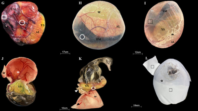
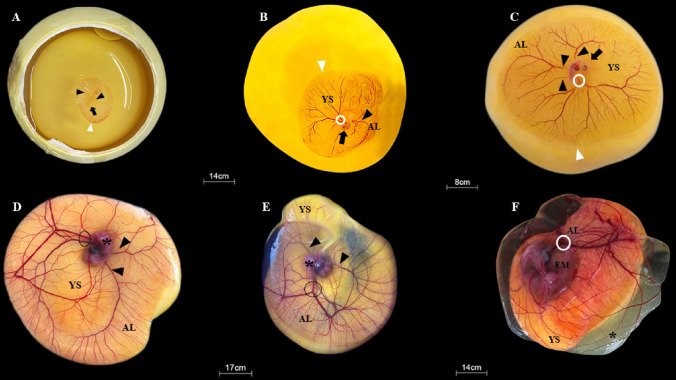
The greater rhea, Rhea americana, is a wild ratite of high scientific importance and significant and zootechnical value, especially considering the current development state of Brazilian poultry production, where research aimed at increasing the productivity of these animals has become extremely relevant. Studies concerning fetal attachments and embryonic development are paramount, as they can provide essential information concerning reproductive and nutritional animal management. However, a lack of information on greater rhea fetal morphology is noted. Therefore, the aim of the present study was to establish a standard model for fetal attachments in this species. Greater rhea eggs were incubated from 0 to 36 days, and macroscopic and microscopic embryonic attachment characterizations were performed. Histologically, all embryonic annexes exhibit germ layers, namely the ectoderm (outer layer), mesoderm (middle layer) and endoderm (inner layer). The findings indicate that greater rhea development patterns are similar to other birds.
期刊介绍:
The journal publishes original research papers, reviews and method papers. While reviews should be designed as comparative surveys, summarizing the current knowledge from an evolutionary perspective, method papers should present new approaches or reviews on methods used in animal morphology. The research papers should be based on morphological investigation of invertebrates and vertebrates at the macroscopic, microscopic and ultrastructural level, including embryological studies.

 求助内容:
求助内容: 应助结果提醒方式:


