Belma Kayhan, Şükrü Sevinçli, Nur Demir, Serkan Demir, Murat Sönmez
{"title":"伴与不伴视神经炎的多发性硬化症视网膜内层改变的区域分析。","authors":"Belma Kayhan, Şükrü Sevinçli, Nur Demir, Serkan Demir, Murat Sönmez","doi":"10.4274/tjo.galenos.2023.81486","DOIUrl":null,"url":null,"abstract":"<p><strong>Objectives: </strong>The study aimed to investigate inner retinal changes in multiple sclerosis (MS) patients by comparing them with healthy controls. The study also aimed to assess regional differences of inner retinal layer involvement in eyes with and without optic neuritis (ON).</p><p><strong>Materials and methods: </strong>This retrospective, cross-sectional study consisted of 141 eyes of 74 relapsing-remitting MS patients and 80 eyes of 40 healthy controls. The study group was separated into two subgroups according to the presence of ON history. Peripapillary retinal nerve fiber layer (pRNFL) thickness, total macular thickness, and thicknesses of the macular retinal nerve fiber layer (mRNFL), ganglion cell layer (GCL), inner plexiform layer (IPL), and inner nuclear layer were compared between the MS and healthy control groups and between eyes with and without ON history.</p><p><strong>Results: </strong>Mean pRNFL, total macular, mRNFL, GCL, and IPL thicknesses were significantly thinner in the MS group than in the control group (p<0.001) and in eyes with ON compared to those without ON (p<0.05). Comparison of inner retinal layer thicknesses in the inner 3-mm ring subfields of the ETDRS grid revealed significant thinning in all subfields of the GCL and IPL of eyes with ON (p<0.05). The inferior subfield demonstrated the highest difference.</p><p><strong>Conclusion: </strong>The study demonstrated that GCL and IPL thinning is a robust and reliable biomarker in all MS patients. The thinning was significantly greater in eyes with ON than in eyes without ON. The study also documented that the inferior region showed significantly greater GCL and IPL thinning in eyes with previous ON attacks.</p>","PeriodicalId":23373,"journal":{"name":"Turkish Journal of Ophthalmology","volume":"53 3","pages":"169-174"},"PeriodicalIF":0.0000,"publicationDate":"2023-06-21","publicationTypes":"Journal Article","fieldsOfStudy":null,"isOpenAccess":false,"openAccessPdf":"https://ftp.ncbi.nlm.nih.gov/pub/pmc/oa_pdf/d4/1d/TJO-53-169.PMC10286840.pdf","citationCount":"0","resultStr":"{\"title\":\"Regional Analysis of Inner Retinal Layer Changes in Multiple Sclerosis with and without Optic Neuritis.\",\"authors\":\"Belma Kayhan, Şükrü Sevinçli, Nur Demir, Serkan Demir, Murat Sönmez\",\"doi\":\"10.4274/tjo.galenos.2023.81486\",\"DOIUrl\":null,\"url\":null,\"abstract\":\"<p><strong>Objectives: </strong>The study aimed to investigate inner retinal changes in multiple sclerosis (MS) patients by comparing them with healthy controls. The study also aimed to assess regional differences of inner retinal layer involvement in eyes with and without optic neuritis (ON).</p><p><strong>Materials and methods: </strong>This retrospective, cross-sectional study consisted of 141 eyes of 74 relapsing-remitting MS patients and 80 eyes of 40 healthy controls. The study group was separated into two subgroups according to the presence of ON history. Peripapillary retinal nerve fiber layer (pRNFL) thickness, total macular thickness, and thicknesses of the macular retinal nerve fiber layer (mRNFL), ganglion cell layer (GCL), inner plexiform layer (IPL), and inner nuclear layer were compared between the MS and healthy control groups and between eyes with and without ON history.</p><p><strong>Results: </strong>Mean pRNFL, total macular, mRNFL, GCL, and IPL thicknesses were significantly thinner in the MS group than in the control group (p<0.001) and in eyes with ON compared to those without ON (p<0.05). Comparison of inner retinal layer thicknesses in the inner 3-mm ring subfields of the ETDRS grid revealed significant thinning in all subfields of the GCL and IPL of eyes with ON (p<0.05). The inferior subfield demonstrated the highest difference.</p><p><strong>Conclusion: </strong>The study demonstrated that GCL and IPL thinning is a robust and reliable biomarker in all MS patients. The thinning was significantly greater in eyes with ON than in eyes without ON. The study also documented that the inferior region showed significantly greater GCL and IPL thinning in eyes with previous ON attacks.</p>\",\"PeriodicalId\":23373,\"journal\":{\"name\":\"Turkish Journal of Ophthalmology\",\"volume\":\"53 3\",\"pages\":\"169-174\"},\"PeriodicalIF\":0.0000,\"publicationDate\":\"2023-06-21\",\"publicationTypes\":\"Journal Article\",\"fieldsOfStudy\":null,\"isOpenAccess\":false,\"openAccessPdf\":\"https://ftp.ncbi.nlm.nih.gov/pub/pmc/oa_pdf/d4/1d/TJO-53-169.PMC10286840.pdf\",\"citationCount\":\"0\",\"resultStr\":null,\"platform\":\"Semanticscholar\",\"paperid\":null,\"PeriodicalName\":\"Turkish Journal of Ophthalmology\",\"FirstCategoryId\":\"1085\",\"ListUrlMain\":\"https://doi.org/10.4274/tjo.galenos.2023.81486\",\"RegionNum\":0,\"RegionCategory\":null,\"ArticlePicture\":[],\"TitleCN\":null,\"AbstractTextCN\":null,\"PMCID\":null,\"EPubDate\":\"\",\"PubModel\":\"\",\"JCR\":\"Q3\",\"JCRName\":\"Medicine\",\"Score\":null,\"Total\":0}","platform":"Semanticscholar","paperid":null,"PeriodicalName":"Turkish Journal of Ophthalmology","FirstCategoryId":"1085","ListUrlMain":"https://doi.org/10.4274/tjo.galenos.2023.81486","RegionNum":0,"RegionCategory":null,"ArticlePicture":[],"TitleCN":null,"AbstractTextCN":null,"PMCID":null,"EPubDate":"","PubModel":"","JCR":"Q3","JCRName":"Medicine","Score":null,"Total":0}
Regional Analysis of Inner Retinal Layer Changes in Multiple Sclerosis with and without Optic Neuritis.
Objectives: The study aimed to investigate inner retinal changes in multiple sclerosis (MS) patients by comparing them with healthy controls. The study also aimed to assess regional differences of inner retinal layer involvement in eyes with and without optic neuritis (ON).
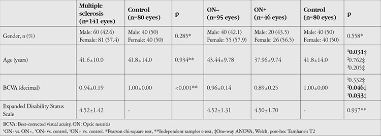
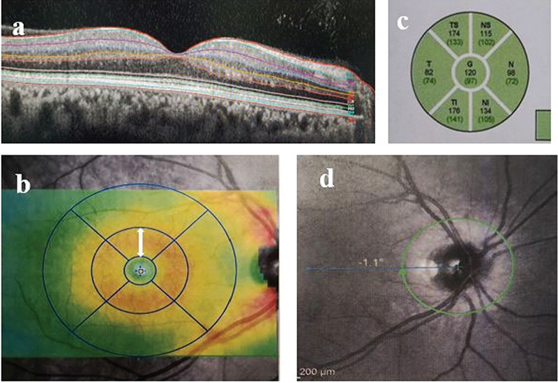
Materials and methods: This retrospective, cross-sectional study consisted of 141 eyes of 74 relapsing-remitting MS patients and 80 eyes of 40 healthy controls. The study group was separated into two subgroups according to the presence of ON history. Peripapillary retinal nerve fiber layer (pRNFL) thickness, total macular thickness, and thicknesses of the macular retinal nerve fiber layer (mRNFL), ganglion cell layer (GCL), inner plexiform layer (IPL), and inner nuclear layer were compared between the MS and healthy control groups and between eyes with and without ON history.
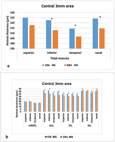
Results: Mean pRNFL, total macular, mRNFL, GCL, and IPL thicknesses were significantly thinner in the MS group than in the control group (p<0.001) and in eyes with ON compared to those without ON (p<0.05). Comparison of inner retinal layer thicknesses in the inner 3-mm ring subfields of the ETDRS grid revealed significant thinning in all subfields of the GCL and IPL of eyes with ON (p<0.05). The inferior subfield demonstrated the highest difference.
Conclusion: The study demonstrated that GCL and IPL thinning is a robust and reliable biomarker in all MS patients. The thinning was significantly greater in eyes with ON than in eyes without ON. The study also documented that the inferior region showed significantly greater GCL and IPL thinning in eyes with previous ON attacks.
期刊介绍:
The Turkish Journal of Ophthalmology (TJO) is the only scientific periodical publication of the Turkish Ophthalmological Association and has been published since January 1929. In its early years, the journal was published in Turkish and French. Although there were temporary interruptions in the publication of the journal due to various challenges, the Turkish Journal of Ophthalmology has been published continually from 1971 to the present. The target audience includes specialists and physicians in training in ophthalmology in all relevant disciplines.

 求助内容:
求助内容: 应助结果提醒方式:


