Sirel Gür Güngör, Şefik Cezairlioğlu, Ahmet Akman, Ümit Ekşioğlu, Almila Sarıgül Sezenöz, Meriç Yavuz Çolak
{"title":"单侧青光眼患者非青光眼的黄斑和乳头周围血管密度。","authors":"Sirel Gür Güngör, Şefik Cezairlioğlu, Ahmet Akman, Ümit Ekşioğlu, Almila Sarıgül Sezenöz, Meriç Yavuz Çolak","doi":"10.4274/tjo.galenos.2022.68302","DOIUrl":null,"url":null,"abstract":"<p><strong>Objectives: </strong>Our purpose was to investigate vascular alterations in the non-glaucomatous eyes of patients with unilateral primary open angle glaucoma using optical coherence tomography angiography and to evaluate the role of vascular damage in glaucoma pathogenesis.</p><p><strong>Materials and methods: </strong>This cross-sectional study included 60 eyes of 30 patients with unilateral glaucoma (63.4±8.8 years) and 30 eyes of 30 healthy subjects (65.6±9.1 years). Three groups were formed: group A, affected eyes of unilateral glaucoma patients; Group B, non-glaucomatous eyes of unilateral glaucoma patients; and group C, healthy controls.</p><p><strong>Results: </strong>When group A was compared with groups B and C, significant differences were detected in rim area, cup volume, mean cup/disc ratio, and retinal nerve fiber layer thickness parameters (p<0.001 for all). No significant difference was detected between groups B and C (p>0.05 for all). In peripapillary and macular vessel density (VD) comparisons, all parameters except intradisc VD were found to be lower in group A (p<0.0167 for all). No statistically significant difference was detected between groups B and C (p>0.05 for all).</p><p><strong>Conclusion: </strong>The VD values in eyes with glaucoma were found to be lower than in the other two groups. However, no difference was observed between the non-glaucomatous eyes of glaucoma patients and those of healthy individuals. Thus, the results did not support our hypothesis that VD alterations would be observed in the fellow eyes of patients with unilateral glaucoma if the vascular pathway were responsible in the pathogenesis of glaucoma.</p>","PeriodicalId":23373,"journal":{"name":"Turkish Journal of Ophthalmology","volume":"53 3","pages":"154-160"},"PeriodicalIF":0.0000,"publicationDate":"2023-06-21","publicationTypes":"Journal Article","fieldsOfStudy":null,"isOpenAccess":false,"openAccessPdf":"https://ftp.ncbi.nlm.nih.gov/pub/pmc/oa_pdf/f0/bd/TJO-53-154.PMC10286842.pdf","citationCount":"0","resultStr":"{\"title\":\"Macular and Peripapillary Vascular Densities in Non-Glaucomatous Eyes of Patients with Unilateral Glaucoma.\",\"authors\":\"Sirel Gür Güngör, Şefik Cezairlioğlu, Ahmet Akman, Ümit Ekşioğlu, Almila Sarıgül Sezenöz, Meriç Yavuz Çolak\",\"doi\":\"10.4274/tjo.galenos.2022.68302\",\"DOIUrl\":null,\"url\":null,\"abstract\":\"<p><strong>Objectives: </strong>Our purpose was to investigate vascular alterations in the non-glaucomatous eyes of patients with unilateral primary open angle glaucoma using optical coherence tomography angiography and to evaluate the role of vascular damage in glaucoma pathogenesis.</p><p><strong>Materials and methods: </strong>This cross-sectional study included 60 eyes of 30 patients with unilateral glaucoma (63.4±8.8 years) and 30 eyes of 30 healthy subjects (65.6±9.1 years). Three groups were formed: group A, affected eyes of unilateral glaucoma patients; Group B, non-glaucomatous eyes of unilateral glaucoma patients; and group C, healthy controls.</p><p><strong>Results: </strong>When group A was compared with groups B and C, significant differences were detected in rim area, cup volume, mean cup/disc ratio, and retinal nerve fiber layer thickness parameters (p<0.001 for all). No significant difference was detected between groups B and C (p>0.05 for all). In peripapillary and macular vessel density (VD) comparisons, all parameters except intradisc VD were found to be lower in group A (p<0.0167 for all). No statistically significant difference was detected between groups B and C (p>0.05 for all).</p><p><strong>Conclusion: </strong>The VD values in eyes with glaucoma were found to be lower than in the other two groups. However, no difference was observed between the non-glaucomatous eyes of glaucoma patients and those of healthy individuals. Thus, the results did not support our hypothesis that VD alterations would be observed in the fellow eyes of patients with unilateral glaucoma if the vascular pathway were responsible in the pathogenesis of glaucoma.</p>\",\"PeriodicalId\":23373,\"journal\":{\"name\":\"Turkish Journal of Ophthalmology\",\"volume\":\"53 3\",\"pages\":\"154-160\"},\"PeriodicalIF\":0.0000,\"publicationDate\":\"2023-06-21\",\"publicationTypes\":\"Journal Article\",\"fieldsOfStudy\":null,\"isOpenAccess\":false,\"openAccessPdf\":\"https://ftp.ncbi.nlm.nih.gov/pub/pmc/oa_pdf/f0/bd/TJO-53-154.PMC10286842.pdf\",\"citationCount\":\"0\",\"resultStr\":null,\"platform\":\"Semanticscholar\",\"paperid\":null,\"PeriodicalName\":\"Turkish Journal of Ophthalmology\",\"FirstCategoryId\":\"1085\",\"ListUrlMain\":\"https://doi.org/10.4274/tjo.galenos.2022.68302\",\"RegionNum\":0,\"RegionCategory\":null,\"ArticlePicture\":[],\"TitleCN\":null,\"AbstractTextCN\":null,\"PMCID\":null,\"EPubDate\":\"\",\"PubModel\":\"\",\"JCR\":\"Q3\",\"JCRName\":\"Medicine\",\"Score\":null,\"Total\":0}","platform":"Semanticscholar","paperid":null,"PeriodicalName":"Turkish Journal of Ophthalmology","FirstCategoryId":"1085","ListUrlMain":"https://doi.org/10.4274/tjo.galenos.2022.68302","RegionNum":0,"RegionCategory":null,"ArticlePicture":[],"TitleCN":null,"AbstractTextCN":null,"PMCID":null,"EPubDate":"","PubModel":"","JCR":"Q3","JCRName":"Medicine","Score":null,"Total":0}
Macular and Peripapillary Vascular Densities in Non-Glaucomatous Eyes of Patients with Unilateral Glaucoma.
Objectives: Our purpose was to investigate vascular alterations in the non-glaucomatous eyes of patients with unilateral primary open angle glaucoma using optical coherence tomography angiography and to evaluate the role of vascular damage in glaucoma pathogenesis.
Materials and methods: This cross-sectional study included 60 eyes of 30 patients with unilateral glaucoma (63.4±8.8 years) and 30 eyes of 30 healthy subjects (65.6±9.1 years). Three groups were formed: group A, affected eyes of unilateral glaucoma patients; Group B, non-glaucomatous eyes of unilateral glaucoma patients; and group C, healthy controls.
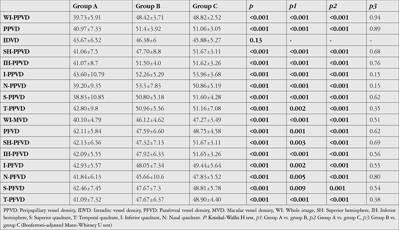
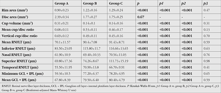
Results: When group A was compared with groups B and C, significant differences were detected in rim area, cup volume, mean cup/disc ratio, and retinal nerve fiber layer thickness parameters (p<0.001 for all). No significant difference was detected between groups B and C (p>0.05 for all). In peripapillary and macular vessel density (VD) comparisons, all parameters except intradisc VD were found to be lower in group A (p<0.0167 for all). No statistically significant difference was detected between groups B and C (p>0.05 for all).
Conclusion: The VD values in eyes with glaucoma were found to be lower than in the other two groups. However, no difference was observed between the non-glaucomatous eyes of glaucoma patients and those of healthy individuals. Thus, the results did not support our hypothesis that VD alterations would be observed in the fellow eyes of patients with unilateral glaucoma if the vascular pathway were responsible in the pathogenesis of glaucoma.
期刊介绍:
The Turkish Journal of Ophthalmology (TJO) is the only scientific periodical publication of the Turkish Ophthalmological Association and has been published since January 1929. In its early years, the journal was published in Turkish and French. Although there were temporary interruptions in the publication of the journal due to various challenges, the Turkish Journal of Ophthalmology has been published continually from 1971 to the present. The target audience includes specialists and physicians in training in ophthalmology in all relevant disciplines.


 求助内容:
求助内容: 应助结果提醒方式:


