Aikaterini Theodorou, Athanasios Tsibonakis, Ioannis S Pateras, Georgia Kaloudi, Eleni Bakola, Maria Chondrogianni, Elissavet Andreadou, Ioannis G Panayiotides, Georgios Tsivgoulis
{"title":"多发性脑微梗死:脑淀粉样血管病相关炎症的罕见表现。","authors":"Aikaterini Theodorou, Athanasios Tsibonakis, Ioannis S Pateras, Georgia Kaloudi, Eleni Bakola, Maria Chondrogianni, Elissavet Andreadou, Ioannis G Panayiotides, Georgios Tsivgoulis","doi":"10.1186/s42466-023-00253-9","DOIUrl":null,"url":null,"abstract":"<p><strong>Background: </strong>Cerebral Amyloid Angiopathy-related inflammation (CAA-ri) is a distinct but rare subset of CAA. The greater availability of high resolution Magnetic Resonance Imaging (MRI) has currently allowed the increasing recognition and diagnosis of this entity, without the risk of a brain biopsy. However, in rare cases with typical clinical characteristics but uncommon neuroimaging findings at presentation, the brain-biopsy is required for an early and reliable diagnosis.</p><p><strong>Case description: </strong>A 71-year-old man with arterial hypertension presented due to 1-week history of headache, vomiting, disorientation and impaired consciousness. Brain MRI revealed multiple acute cortical/subcortical microinfarcts, scarce microbleeds, extensive right parietooccipital and left frontotemporal leptomeningeal enhancement. After an extensive diagnostic work-up, excluding infectious, neoplastic and autoimmune etiologies, the patient underwent brain-biopsy. Histology disclosed amyloid deposition in an arteriolar wall and the patient fulfilled diagnostic criteria for probable CAA-ri with supporting pathology. He received intravenous methylprednisolone, followed by oral tapering with steroids showing clinical and radiological improvement with complete resolution of gadolinium enhancement. Follow-up MRI revealed an increase of cerebral microbleeds and the patient fulfilled CAA-ri neuroimaging criteria.</p><p><strong>Conclusions: </strong>This case highlights the importance of continuous vigilance from clinical neurologists to detect CAA-ri diagnosis and the diagnostic value of brain-biopsy in CAA-ri patients with atypical neuroimaging presentation, such as acute microinfarcts. The early diagnosis and the prompt treatment initiation can improve the prognosis and the evolution of this rare disorder.</p>","PeriodicalId":19169,"journal":{"name":"Neurological Research and Practice","volume":"5 1","pages":"28"},"PeriodicalIF":0.0000,"publicationDate":"2023-06-22","publicationTypes":"Journal Article","fieldsOfStudy":null,"isOpenAccess":false,"openAccessPdf":"https://www.ncbi.nlm.nih.gov/pmc/articles/PMC10286471/pdf/","citationCount":"0","resultStr":"{\"title\":\"Multiple cerebral microinfarcts: an uncommon presentation of Cerebral Amyloid Angiopathy-related inflammation.\",\"authors\":\"Aikaterini Theodorou, Athanasios Tsibonakis, Ioannis S Pateras, Georgia Kaloudi, Eleni Bakola, Maria Chondrogianni, Elissavet Andreadou, Ioannis G Panayiotides, Georgios Tsivgoulis\",\"doi\":\"10.1186/s42466-023-00253-9\",\"DOIUrl\":null,\"url\":null,\"abstract\":\"<p><strong>Background: </strong>Cerebral Amyloid Angiopathy-related inflammation (CAA-ri) is a distinct but rare subset of CAA. The greater availability of high resolution Magnetic Resonance Imaging (MRI) has currently allowed the increasing recognition and diagnosis of this entity, without the risk of a brain biopsy. However, in rare cases with typical clinical characteristics but uncommon neuroimaging findings at presentation, the brain-biopsy is required for an early and reliable diagnosis.</p><p><strong>Case description: </strong>A 71-year-old man with arterial hypertension presented due to 1-week history of headache, vomiting, disorientation and impaired consciousness. Brain MRI revealed multiple acute cortical/subcortical microinfarcts, scarce microbleeds, extensive right parietooccipital and left frontotemporal leptomeningeal enhancement. After an extensive diagnostic work-up, excluding infectious, neoplastic and autoimmune etiologies, the patient underwent brain-biopsy. Histology disclosed amyloid deposition in an arteriolar wall and the patient fulfilled diagnostic criteria for probable CAA-ri with supporting pathology. He received intravenous methylprednisolone, followed by oral tapering with steroids showing clinical and radiological improvement with complete resolution of gadolinium enhancement. Follow-up MRI revealed an increase of cerebral microbleeds and the patient fulfilled CAA-ri neuroimaging criteria.</p><p><strong>Conclusions: </strong>This case highlights the importance of continuous vigilance from clinical neurologists to detect CAA-ri diagnosis and the diagnostic value of brain-biopsy in CAA-ri patients with atypical neuroimaging presentation, such as acute microinfarcts. The early diagnosis and the prompt treatment initiation can improve the prognosis and the evolution of this rare disorder.</p>\",\"PeriodicalId\":19169,\"journal\":{\"name\":\"Neurological Research and Practice\",\"volume\":\"5 1\",\"pages\":\"28\"},\"PeriodicalIF\":0.0000,\"publicationDate\":\"2023-06-22\",\"publicationTypes\":\"Journal Article\",\"fieldsOfStudy\":null,\"isOpenAccess\":false,\"openAccessPdf\":\"https://www.ncbi.nlm.nih.gov/pmc/articles/PMC10286471/pdf/\",\"citationCount\":\"0\",\"resultStr\":null,\"platform\":\"Semanticscholar\",\"paperid\":null,\"PeriodicalName\":\"Neurological Research and Practice\",\"FirstCategoryId\":\"1085\",\"ListUrlMain\":\"https://doi.org/10.1186/s42466-023-00253-9\",\"RegionNum\":0,\"RegionCategory\":null,\"ArticlePicture\":[],\"TitleCN\":null,\"AbstractTextCN\":null,\"PMCID\":null,\"EPubDate\":\"\",\"PubModel\":\"\",\"JCR\":\"\",\"JCRName\":\"\",\"Score\":null,\"Total\":0}","platform":"Semanticscholar","paperid":null,"PeriodicalName":"Neurological Research and Practice","FirstCategoryId":"1085","ListUrlMain":"https://doi.org/10.1186/s42466-023-00253-9","RegionNum":0,"RegionCategory":null,"ArticlePicture":[],"TitleCN":null,"AbstractTextCN":null,"PMCID":null,"EPubDate":"","PubModel":"","JCR":"","JCRName":"","Score":null,"Total":0}
Multiple cerebral microinfarcts: an uncommon presentation of Cerebral Amyloid Angiopathy-related inflammation.
Background: Cerebral Amyloid Angiopathy-related inflammation (CAA-ri) is a distinct but rare subset of CAA. The greater availability of high resolution Magnetic Resonance Imaging (MRI) has currently allowed the increasing recognition and diagnosis of this entity, without the risk of a brain biopsy. However, in rare cases with typical clinical characteristics but uncommon neuroimaging findings at presentation, the brain-biopsy is required for an early and reliable diagnosis.
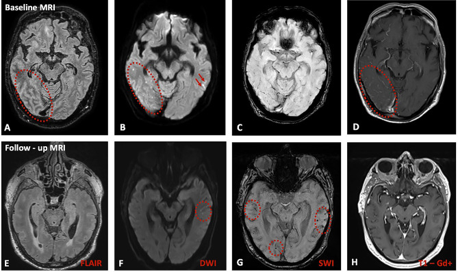
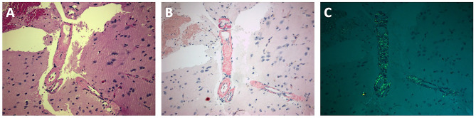
Case description: A 71-year-old man with arterial hypertension presented due to 1-week history of headache, vomiting, disorientation and impaired consciousness. Brain MRI revealed multiple acute cortical/subcortical microinfarcts, scarce microbleeds, extensive right parietooccipital and left frontotemporal leptomeningeal enhancement. After an extensive diagnostic work-up, excluding infectious, neoplastic and autoimmune etiologies, the patient underwent brain-biopsy. Histology disclosed amyloid deposition in an arteriolar wall and the patient fulfilled diagnostic criteria for probable CAA-ri with supporting pathology. He received intravenous methylprednisolone, followed by oral tapering with steroids showing clinical and radiological improvement with complete resolution of gadolinium enhancement. Follow-up MRI revealed an increase of cerebral microbleeds and the patient fulfilled CAA-ri neuroimaging criteria.
Conclusions: This case highlights the importance of continuous vigilance from clinical neurologists to detect CAA-ri diagnosis and the diagnostic value of brain-biopsy in CAA-ri patients with atypical neuroimaging presentation, such as acute microinfarcts. The early diagnosis and the prompt treatment initiation can improve the prognosis and the evolution of this rare disorder.

 求助内容:
求助内容: 应助结果提醒方式:


