{"title":"立体定向放射外科治疗后白色表皮样囊肿的转化:一例说明。","authors":"Hideki Matsumoto, Yuki Shinya, Satoru Miyawaki, Masahiro Shin, Satoshi Koizumi, Daisuke Sato, Munetoshi Hinata, Masako Ikemura, Satoshi Kiyofuji, Taich Kin, Mototaro Iwanaga, Masahiro Shimizu, Hirofumi Nakatomi, Nobuhito Saito","doi":"10.3171/CASE2376","DOIUrl":null,"url":null,"abstract":"<p><strong>Background: </strong>White epidermoid cysts (WECs) are a rare type of epidermoid cyst with atypical radiological features. The epidemiological aspects and mechanisms of their onset remain unknown. Herein, the authors report a unique case of WEC transformation from a typical epidermoid cyst after stereotactic radiosurgery (SRS), confirmed by radiological and pathological findings.</p><p><strong>Observations: </strong>The case involved a 78-year-old man with a history of 2 surgeries for a left cerebellopontine angle typical epidermoid cyst 23 years earlier and SRS using the CyberKnife for recurrent trigeminal neuralgia (TN) 14 years earlier. The tumor with high intensity on T1-weighted imaging, low intensity on T2-weighted imaging, without restriction on diffusion-weighted imaging had gradually enlarged after SRS. Therefore, a salvage surgery was performed via a left suboccipital craniotomy, and the intraoperative findings showed a cyst with a brown, viscous liquid component, consistent with those of WECs. Histopathologically, keratin calcification and hemorrhage were identified, leading to a diagnosis of WEC. The postoperative course was uneventful, and the TN resolved. No tumor recurrence was recorded at 2 years postoperatively.</p><p><strong>Lessons: </strong>To the best of the authors' knowledge, this is the first world case of WEC transformation from a typical epidermoid cyst after SRS, confirmed by radiological and pathological findings. Radiation effects could have been involved in this transformation.</p>","PeriodicalId":16554,"journal":{"name":"Journal of Neurosurgery: Case Lessons","volume":"5 24","pages":""},"PeriodicalIF":0.0000,"publicationDate":"2023-06-12","publicationTypes":"Journal Article","fieldsOfStudy":null,"isOpenAccess":false,"openAccessPdf":"https://ftp.ncbi.nlm.nih.gov/pub/pmc/oa_pdf/77/a5/CASE2376.PMC10550655.pdf","citationCount":"0","resultStr":"{\"title\":\"White epidermoid cyst transformation after stereotactic radiosurgery: illustrative case.\",\"authors\":\"Hideki Matsumoto, Yuki Shinya, Satoru Miyawaki, Masahiro Shin, Satoshi Koizumi, Daisuke Sato, Munetoshi Hinata, Masako Ikemura, Satoshi Kiyofuji, Taich Kin, Mototaro Iwanaga, Masahiro Shimizu, Hirofumi Nakatomi, Nobuhito Saito\",\"doi\":\"10.3171/CASE2376\",\"DOIUrl\":null,\"url\":null,\"abstract\":\"<p><strong>Background: </strong>White epidermoid cysts (WECs) are a rare type of epidermoid cyst with atypical radiological features. The epidemiological aspects and mechanisms of their onset remain unknown. Herein, the authors report a unique case of WEC transformation from a typical epidermoid cyst after stereotactic radiosurgery (SRS), confirmed by radiological and pathological findings.</p><p><strong>Observations: </strong>The case involved a 78-year-old man with a history of 2 surgeries for a left cerebellopontine angle typical epidermoid cyst 23 years earlier and SRS using the CyberKnife for recurrent trigeminal neuralgia (TN) 14 years earlier. The tumor with high intensity on T1-weighted imaging, low intensity on T2-weighted imaging, without restriction on diffusion-weighted imaging had gradually enlarged after SRS. Therefore, a salvage surgery was performed via a left suboccipital craniotomy, and the intraoperative findings showed a cyst with a brown, viscous liquid component, consistent with those of WECs. Histopathologically, keratin calcification and hemorrhage were identified, leading to a diagnosis of WEC. The postoperative course was uneventful, and the TN resolved. No tumor recurrence was recorded at 2 years postoperatively.</p><p><strong>Lessons: </strong>To the best of the authors' knowledge, this is the first world case of WEC transformation from a typical epidermoid cyst after SRS, confirmed by radiological and pathological findings. Radiation effects could have been involved in this transformation.</p>\",\"PeriodicalId\":16554,\"journal\":{\"name\":\"Journal of Neurosurgery: Case Lessons\",\"volume\":\"5 24\",\"pages\":\"\"},\"PeriodicalIF\":0.0000,\"publicationDate\":\"2023-06-12\",\"publicationTypes\":\"Journal Article\",\"fieldsOfStudy\":null,\"isOpenAccess\":false,\"openAccessPdf\":\"https://ftp.ncbi.nlm.nih.gov/pub/pmc/oa_pdf/77/a5/CASE2376.PMC10550655.pdf\",\"citationCount\":\"0\",\"resultStr\":null,\"platform\":\"Semanticscholar\",\"paperid\":null,\"PeriodicalName\":\"Journal of Neurosurgery: Case Lessons\",\"FirstCategoryId\":\"1085\",\"ListUrlMain\":\"https://doi.org/10.3171/CASE2376\",\"RegionNum\":0,\"RegionCategory\":null,\"ArticlePicture\":[],\"TitleCN\":null,\"AbstractTextCN\":null,\"PMCID\":null,\"EPubDate\":\"\",\"PubModel\":\"\",\"JCR\":\"\",\"JCRName\":\"\",\"Score\":null,\"Total\":0}","platform":"Semanticscholar","paperid":null,"PeriodicalName":"Journal of Neurosurgery: Case Lessons","FirstCategoryId":"1085","ListUrlMain":"https://doi.org/10.3171/CASE2376","RegionNum":0,"RegionCategory":null,"ArticlePicture":[],"TitleCN":null,"AbstractTextCN":null,"PMCID":null,"EPubDate":"","PubModel":"","JCR":"","JCRName":"","Score":null,"Total":0}
White epidermoid cyst transformation after stereotactic radiosurgery: illustrative case.
Background: White epidermoid cysts (WECs) are a rare type of epidermoid cyst with atypical radiological features. The epidemiological aspects and mechanisms of their onset remain unknown. Herein, the authors report a unique case of WEC transformation from a typical epidermoid cyst after stereotactic radiosurgery (SRS), confirmed by radiological and pathological findings.
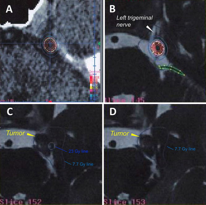
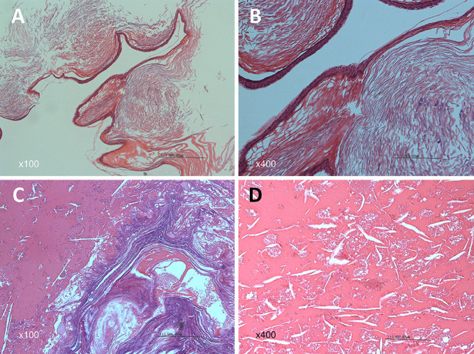
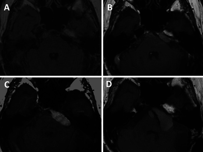
Observations: The case involved a 78-year-old man with a history of 2 surgeries for a left cerebellopontine angle typical epidermoid cyst 23 years earlier and SRS using the CyberKnife for recurrent trigeminal neuralgia (TN) 14 years earlier. The tumor with high intensity on T1-weighted imaging, low intensity on T2-weighted imaging, without restriction on diffusion-weighted imaging had gradually enlarged after SRS. Therefore, a salvage surgery was performed via a left suboccipital craniotomy, and the intraoperative findings showed a cyst with a brown, viscous liquid component, consistent with those of WECs. Histopathologically, keratin calcification and hemorrhage were identified, leading to a diagnosis of WEC. The postoperative course was uneventful, and the TN resolved. No tumor recurrence was recorded at 2 years postoperatively.
Lessons: To the best of the authors' knowledge, this is the first world case of WEC transformation from a typical epidermoid cyst after SRS, confirmed by radiological and pathological findings. Radiation effects could have been involved in this transformation.

 求助内容:
求助内容: 应助结果提醒方式:


