Seung Jun Lee, Youe Ree Kim, Young Hwan Lee, Kwon-Ha Yoon
{"title":"超声衰减成像对脂肪肝的评价和诊断。","authors":"Seung Jun Lee, Youe Ree Kim, Young Hwan Lee, Kwon-Ha Yoon","doi":"10.3348/jksr.2022.0053","DOIUrl":null,"url":null,"abstract":"<p><strong>Purpose: </strong>This study aimed to determine whether the attenuation coefficient (AC) from attenuation imaging (ATI) was correlated with visual US assessment in patients with hepatic steatosis. Moreover, it aimed to assess whether the patient's blood chemistry results and CT attenuation were correlated with AC.</p><p><strong>Materials and methods: </strong>Patients who underwent abdominal US with ATI between April 2018 and December 2018 were included in this study. Patients with chronic liver disease or cirrhosis were excluded. The correlation between AC and other parameters, such as visual US assessment, blood chemistry results, liver attenuation, and liver-to-spleen (L/S) ratio, were analyzed. AC values according to visual US assessment grades were compared using analysis of variance.</p><p><strong>Results: </strong>A total of 161 patients were included in this study. The correlation coefficient between US assessment and AC was 0.814 (<i>p</i> < 0.001). The mean AC values for the normal, mild, moderate, and severe grades were 0.56, 0.66, 0.74, and 0.85, respectively (<i>p</i> < 0.001). Alanine aminotransferase levels were significantly correlated with AC (<i>r</i> = 0.317, <i>p</i> < 0.001). The correlation coefficients between liver attenuation and AC and between L/S ratio and AC were -0.702 and -0.626, respectively (<i>p</i> < 0.001).</p><p><strong>Conclusion: </strong>Visual US assessment and AC showed a strong positive correlation with the discriminative value between the groups. Computed tomography attenuation and AC showed a strong negative correlation.</p>","PeriodicalId":17455,"journal":{"name":"Journal of the Korean Society of Radiology","volume":"84 3","pages":"666-675"},"PeriodicalIF":0.0000,"publicationDate":"2023-05-01","publicationTypes":"Journal Article","fieldsOfStudy":null,"isOpenAccess":false,"openAccessPdf":"https://ftp.ncbi.nlm.nih.gov/pub/pmc/oa_pdf/d9/2a/jksr-84-666.PMC10265227.pdf","citationCount":"0","resultStr":"{\"title\":\"US Attenuation Imaging for the Evaluation and Diagnosis of Fatty Liver Disease.\",\"authors\":\"Seung Jun Lee, Youe Ree Kim, Young Hwan Lee, Kwon-Ha Yoon\",\"doi\":\"10.3348/jksr.2022.0053\",\"DOIUrl\":null,\"url\":null,\"abstract\":\"<p><strong>Purpose: </strong>This study aimed to determine whether the attenuation coefficient (AC) from attenuation imaging (ATI) was correlated with visual US assessment in patients with hepatic steatosis. Moreover, it aimed to assess whether the patient's blood chemistry results and CT attenuation were correlated with AC.</p><p><strong>Materials and methods: </strong>Patients who underwent abdominal US with ATI between April 2018 and December 2018 were included in this study. Patients with chronic liver disease or cirrhosis were excluded. The correlation between AC and other parameters, such as visual US assessment, blood chemistry results, liver attenuation, and liver-to-spleen (L/S) ratio, were analyzed. AC values according to visual US assessment grades were compared using analysis of variance.</p><p><strong>Results: </strong>A total of 161 patients were included in this study. The correlation coefficient between US assessment and AC was 0.814 (<i>p</i> < 0.001). The mean AC values for the normal, mild, moderate, and severe grades were 0.56, 0.66, 0.74, and 0.85, respectively (<i>p</i> < 0.001). Alanine aminotransferase levels were significantly correlated with AC (<i>r</i> = 0.317, <i>p</i> < 0.001). The correlation coefficients between liver attenuation and AC and between L/S ratio and AC were -0.702 and -0.626, respectively (<i>p</i> < 0.001).</p><p><strong>Conclusion: </strong>Visual US assessment and AC showed a strong positive correlation with the discriminative value between the groups. Computed tomography attenuation and AC showed a strong negative correlation.</p>\",\"PeriodicalId\":17455,\"journal\":{\"name\":\"Journal of the Korean Society of Radiology\",\"volume\":\"84 3\",\"pages\":\"666-675\"},\"PeriodicalIF\":0.0000,\"publicationDate\":\"2023-05-01\",\"publicationTypes\":\"Journal Article\",\"fieldsOfStudy\":null,\"isOpenAccess\":false,\"openAccessPdf\":\"https://ftp.ncbi.nlm.nih.gov/pub/pmc/oa_pdf/d9/2a/jksr-84-666.PMC10265227.pdf\",\"citationCount\":\"0\",\"resultStr\":null,\"platform\":\"Semanticscholar\",\"paperid\":null,\"PeriodicalName\":\"Journal of the Korean Society of Radiology\",\"FirstCategoryId\":\"1085\",\"ListUrlMain\":\"https://doi.org/10.3348/jksr.2022.0053\",\"RegionNum\":0,\"RegionCategory\":null,\"ArticlePicture\":[],\"TitleCN\":null,\"AbstractTextCN\":null,\"PMCID\":null,\"EPubDate\":\"\",\"PubModel\":\"\",\"JCR\":\"Q4\",\"JCRName\":\"Medicine\",\"Score\":null,\"Total\":0}","platform":"Semanticscholar","paperid":null,"PeriodicalName":"Journal of the Korean Society of Radiology","FirstCategoryId":"1085","ListUrlMain":"https://doi.org/10.3348/jksr.2022.0053","RegionNum":0,"RegionCategory":null,"ArticlePicture":[],"TitleCN":null,"AbstractTextCN":null,"PMCID":null,"EPubDate":"","PubModel":"","JCR":"Q4","JCRName":"Medicine","Score":null,"Total":0}
US Attenuation Imaging for the Evaluation and Diagnosis of Fatty Liver Disease.
Purpose: This study aimed to determine whether the attenuation coefficient (AC) from attenuation imaging (ATI) was correlated with visual US assessment in patients with hepatic steatosis. Moreover, it aimed to assess whether the patient's blood chemistry results and CT attenuation were correlated with AC.
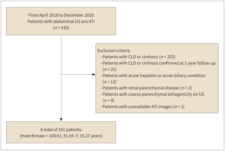
Materials and methods: Patients who underwent abdominal US with ATI between April 2018 and December 2018 were included in this study. Patients with chronic liver disease or cirrhosis were excluded. The correlation between AC and other parameters, such as visual US assessment, blood chemistry results, liver attenuation, and liver-to-spleen (L/S) ratio, were analyzed. AC values according to visual US assessment grades were compared using analysis of variance.
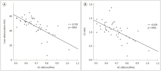
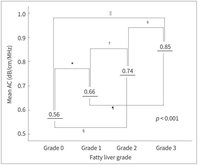
Results: A total of 161 patients were included in this study. The correlation coefficient between US assessment and AC was 0.814 (p < 0.001). The mean AC values for the normal, mild, moderate, and severe grades were 0.56, 0.66, 0.74, and 0.85, respectively (p < 0.001). Alanine aminotransferase levels were significantly correlated with AC (r = 0.317, p < 0.001). The correlation coefficients between liver attenuation and AC and between L/S ratio and AC were -0.702 and -0.626, respectively (p < 0.001).
Conclusion: Visual US assessment and AC showed a strong positive correlation with the discriminative value between the groups. Computed tomography attenuation and AC showed a strong negative correlation.

 求助内容:
求助内容: 应助结果提醒方式:


