So Ra Shin, Eun Young Ko, Boo-Kyung Han, Eun Sook Ko, Ji Soo Choi, Haejung Kim
{"title":"乳腺良性腺肌瘤:影像学特征。","authors":"So Ra Shin, Eun Young Ko, Boo-Kyung Han, Eun Sook Ko, Ji Soo Choi, Haejung Kim","doi":"10.3348/jksr.2022.0021","DOIUrl":null,"url":null,"abstract":"<p><strong>Purpose: </strong>This study aimed to evaluate the radiological and clinical characteristics of benign adenomyoepitheliomas of the breast.</p><p><strong>Materials and methods: </strong>Over the last 20 years, 120 patients were histologically diagnosed with breast adenomyoepithelioma (AME) at our institution. We excluded 43 patients who were incidentally diagnosed during mastectomy for breast cancer, 28 who underwent percutaneous biopsy without further excision, and 8 who had biopsy-confirmed benign AME and were found to have another pathology after complete excision. We retrospectively reviewed the clinical records and radiological findings of the remaining 41 patients with histologically diagnosed benign breast AMEs after complete excision.</p><p><strong>Results: </strong>All 41 patients underwent US; 38 underwent mammography (MG) and US; and 18 underwent MG, US, and MRI. MG detected 38 cases with a round or oval shape (56%), and mass (89%), were non-circumscribed (62%), hyperdense (53%), and without microcalcifications (95%). Breast US revealed suspicious masses (98%) with a non-circumscribed margin (66%), hypoechogenicity (43%), and intratumoral vascularity (63%). All lesions on breast MRI showed suspicious masses (100%) with ill-defined margins (61%), and 84% showed wash-out kinetics. Benign AMEs showed suspicious features of Breast Imaging Reporting and Data System (BI-RADS) category 4 or 5 in 83%-95% of the MG, US, and MRI. Sixteen of the 41 cases were misdiagnosed on the initial core needle biopsy and two were diagnosed as malignancy.</p><p><strong>Conclusion: </strong>Benign breast AME often shows suspicious radiological features mimicking a malignant mass on MG, US, and MRI. Differentiating benign AME from other pathologies might be difficult on core needle biopsy, and complete excision is needed for a correct diagnosis.</p>","PeriodicalId":17455,"journal":{"name":"Journal of the Korean Society of Radiology","volume":"84 2","pages":"398-408"},"PeriodicalIF":0.0000,"publicationDate":"2023-03-01","publicationTypes":"Journal Article","fieldsOfStudy":null,"isOpenAccess":false,"openAccessPdf":"https://ftp.ncbi.nlm.nih.gov/pub/pmc/oa_pdf/64/97/jksr-84-398.PMC10083621.pdf","citationCount":"0","resultStr":"{\"title\":\"Benign Adenomyoepithelioma of the Breast: Imaging Characteristics.\",\"authors\":\"So Ra Shin, Eun Young Ko, Boo-Kyung Han, Eun Sook Ko, Ji Soo Choi, Haejung Kim\",\"doi\":\"10.3348/jksr.2022.0021\",\"DOIUrl\":null,\"url\":null,\"abstract\":\"<p><strong>Purpose: </strong>This study aimed to evaluate the radiological and clinical characteristics of benign adenomyoepitheliomas of the breast.</p><p><strong>Materials and methods: </strong>Over the last 20 years, 120 patients were histologically diagnosed with breast adenomyoepithelioma (AME) at our institution. We excluded 43 patients who were incidentally diagnosed during mastectomy for breast cancer, 28 who underwent percutaneous biopsy without further excision, and 8 who had biopsy-confirmed benign AME and were found to have another pathology after complete excision. We retrospectively reviewed the clinical records and radiological findings of the remaining 41 patients with histologically diagnosed benign breast AMEs after complete excision.</p><p><strong>Results: </strong>All 41 patients underwent US; 38 underwent mammography (MG) and US; and 18 underwent MG, US, and MRI. MG detected 38 cases with a round or oval shape (56%), and mass (89%), were non-circumscribed (62%), hyperdense (53%), and without microcalcifications (95%). Breast US revealed suspicious masses (98%) with a non-circumscribed margin (66%), hypoechogenicity (43%), and intratumoral vascularity (63%). All lesions on breast MRI showed suspicious masses (100%) with ill-defined margins (61%), and 84% showed wash-out kinetics. Benign AMEs showed suspicious features of Breast Imaging Reporting and Data System (BI-RADS) category 4 or 5 in 83%-95% of the MG, US, and MRI. Sixteen of the 41 cases were misdiagnosed on the initial core needle biopsy and two were diagnosed as malignancy.</p><p><strong>Conclusion: </strong>Benign breast AME often shows suspicious radiological features mimicking a malignant mass on MG, US, and MRI. Differentiating benign AME from other pathologies might be difficult on core needle biopsy, and complete excision is needed for a correct diagnosis.</p>\",\"PeriodicalId\":17455,\"journal\":{\"name\":\"Journal of the Korean Society of Radiology\",\"volume\":\"84 2\",\"pages\":\"398-408\"},\"PeriodicalIF\":0.0000,\"publicationDate\":\"2023-03-01\",\"publicationTypes\":\"Journal Article\",\"fieldsOfStudy\":null,\"isOpenAccess\":false,\"openAccessPdf\":\"https://ftp.ncbi.nlm.nih.gov/pub/pmc/oa_pdf/64/97/jksr-84-398.PMC10083621.pdf\",\"citationCount\":\"0\",\"resultStr\":null,\"platform\":\"Semanticscholar\",\"paperid\":null,\"PeriodicalName\":\"Journal of the Korean Society of Radiology\",\"FirstCategoryId\":\"1085\",\"ListUrlMain\":\"https://doi.org/10.3348/jksr.2022.0021\",\"RegionNum\":0,\"RegionCategory\":null,\"ArticlePicture\":[],\"TitleCN\":null,\"AbstractTextCN\":null,\"PMCID\":null,\"EPubDate\":\"\",\"PubModel\":\"\",\"JCR\":\"Q4\",\"JCRName\":\"Medicine\",\"Score\":null,\"Total\":0}","platform":"Semanticscholar","paperid":null,"PeriodicalName":"Journal of the Korean Society of Radiology","FirstCategoryId":"1085","ListUrlMain":"https://doi.org/10.3348/jksr.2022.0021","RegionNum":0,"RegionCategory":null,"ArticlePicture":[],"TitleCN":null,"AbstractTextCN":null,"PMCID":null,"EPubDate":"","PubModel":"","JCR":"Q4","JCRName":"Medicine","Score":null,"Total":0}
Benign Adenomyoepithelioma of the Breast: Imaging Characteristics.
Purpose: This study aimed to evaluate the radiological and clinical characteristics of benign adenomyoepitheliomas of the breast.
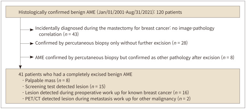
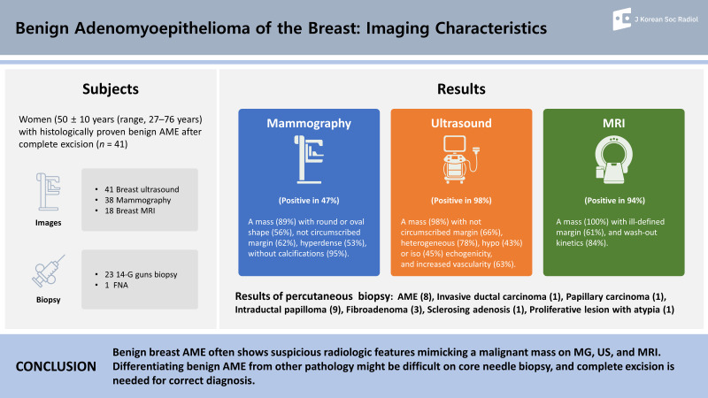
Materials and methods: Over the last 20 years, 120 patients were histologically diagnosed with breast adenomyoepithelioma (AME) at our institution. We excluded 43 patients who were incidentally diagnosed during mastectomy for breast cancer, 28 who underwent percutaneous biopsy without further excision, and 8 who had biopsy-confirmed benign AME and were found to have another pathology after complete excision. We retrospectively reviewed the clinical records and radiological findings of the remaining 41 patients with histologically diagnosed benign breast AMEs after complete excision.
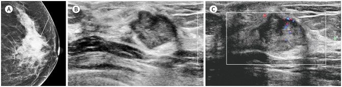
Results: All 41 patients underwent US; 38 underwent mammography (MG) and US; and 18 underwent MG, US, and MRI. MG detected 38 cases with a round or oval shape (56%), and mass (89%), were non-circumscribed (62%), hyperdense (53%), and without microcalcifications (95%). Breast US revealed suspicious masses (98%) with a non-circumscribed margin (66%), hypoechogenicity (43%), and intratumoral vascularity (63%). All lesions on breast MRI showed suspicious masses (100%) with ill-defined margins (61%), and 84% showed wash-out kinetics. Benign AMEs showed suspicious features of Breast Imaging Reporting and Data System (BI-RADS) category 4 or 5 in 83%-95% of the MG, US, and MRI. Sixteen of the 41 cases were misdiagnosed on the initial core needle biopsy and two were diagnosed as malignancy.
Conclusion: Benign breast AME often shows suspicious radiological features mimicking a malignant mass on MG, US, and MRI. Differentiating benign AME from other pathologies might be difficult on core needle biopsy, and complete excision is needed for a correct diagnosis.

 求助内容:
求助内容: 应助结果提醒方式:


