{"title":"正常甲状旁腺的超声特征:与甲状腺癌转移性淋巴结的比较。","authors":"Seong Ju Kim, Dong Gyu Na, Byeong-Joo Noh","doi":"10.14366/usg.22119","DOIUrl":null,"url":null,"abstract":"<p><strong>Purpose: </strong>This study aimed to determine the ultrasound (US) features of normal parathyroid glands (PTGs) and to evaluate whether normal PTGs can be differentiated from metastatic lymph nodes (LNs) in thyroid cancer.</p><p><strong>Methods: </strong>This retrospective study included 10 normal PTGs and 95 metastatic LNs from thyroid cancer showing suspicious US features. The echogenicity, echotexture, echogenic foci (calcifications), cystic change, abnormal vascularity, size, shape, and location were retrospectively assessed and compared between normal PTGs and metastatic LNs.</p><p><strong>Results: </strong>The echogenicity of normal PTGs was significantly different from that of metastatic LNs (P<0.001). Normal PTGs exhibited marked hyperechogenicity (100%), homogeneous echotexture (80%), focal intraglandular hypoechogenicity (20%), ovoid shape (90%), and focal cystic change in one case (10%). The echogenicity of metastatic LNs was markedly hyperechoic (0%), moderately hyperechoic (15.8%), mildly hyperechoic (53.7%), and hypoechoic (28.4%). The size and long axis/short axis ratios of normal PTGs were significantly smaller and larger than those of metastatic LNs (P<0.01 and P=0.022, respectively).</p><p><strong>Conclusion: </strong>Marked hyperechogenicity was found only in normal PTGs, and small, ovoid, markedly hyperechoic structures in the paramedian central neck characterized normal PTGs. Normal PTGs may be differentiated from metastatic LNs in thyroid cancer.</p>","PeriodicalId":54227,"journal":{"name":"Ultrasonography","volume":"42 2","pages":"203-213"},"PeriodicalIF":2.4000,"publicationDate":"2023-04-01","publicationTypes":"Journal Article","fieldsOfStudy":null,"isOpenAccess":false,"openAccessPdf":"https://ftp.ncbi.nlm.nih.gov/pub/pmc/oa_pdf/be/b6/usg-22119.PMC10071065.pdf","citationCount":"1","resultStr":"{\"title\":\"US features of normal parathyroid glands: a comparison with metastatic lymph nodes in thyroid cancer.\",\"authors\":\"Seong Ju Kim, Dong Gyu Na, Byeong-Joo Noh\",\"doi\":\"10.14366/usg.22119\",\"DOIUrl\":null,\"url\":null,\"abstract\":\"<p><strong>Purpose: </strong>This study aimed to determine the ultrasound (US) features of normal parathyroid glands (PTGs) and to evaluate whether normal PTGs can be differentiated from metastatic lymph nodes (LNs) in thyroid cancer.</p><p><strong>Methods: </strong>This retrospective study included 10 normal PTGs and 95 metastatic LNs from thyroid cancer showing suspicious US features. The echogenicity, echotexture, echogenic foci (calcifications), cystic change, abnormal vascularity, size, shape, and location were retrospectively assessed and compared between normal PTGs and metastatic LNs.</p><p><strong>Results: </strong>The echogenicity of normal PTGs was significantly different from that of metastatic LNs (P<0.001). Normal PTGs exhibited marked hyperechogenicity (100%), homogeneous echotexture (80%), focal intraglandular hypoechogenicity (20%), ovoid shape (90%), and focal cystic change in one case (10%). The echogenicity of metastatic LNs was markedly hyperechoic (0%), moderately hyperechoic (15.8%), mildly hyperechoic (53.7%), and hypoechoic (28.4%). The size and long axis/short axis ratios of normal PTGs were significantly smaller and larger than those of metastatic LNs (P<0.01 and P=0.022, respectively).</p><p><strong>Conclusion: </strong>Marked hyperechogenicity was found only in normal PTGs, and small, ovoid, markedly hyperechoic structures in the paramedian central neck characterized normal PTGs. Normal PTGs may be differentiated from metastatic LNs in thyroid cancer.</p>\",\"PeriodicalId\":54227,\"journal\":{\"name\":\"Ultrasonography\",\"volume\":\"42 2\",\"pages\":\"203-213\"},\"PeriodicalIF\":2.4000,\"publicationDate\":\"2023-04-01\",\"publicationTypes\":\"Journal Article\",\"fieldsOfStudy\":null,\"isOpenAccess\":false,\"openAccessPdf\":\"https://ftp.ncbi.nlm.nih.gov/pub/pmc/oa_pdf/be/b6/usg-22119.PMC10071065.pdf\",\"citationCount\":\"1\",\"resultStr\":null,\"platform\":\"Semanticscholar\",\"paperid\":null,\"PeriodicalName\":\"Ultrasonography\",\"FirstCategoryId\":\"3\",\"ListUrlMain\":\"https://doi.org/10.14366/usg.22119\",\"RegionNum\":3,\"RegionCategory\":\"医学\",\"ArticlePicture\":[],\"TitleCN\":null,\"AbstractTextCN\":null,\"PMCID\":null,\"EPubDate\":\"\",\"PubModel\":\"\",\"JCR\":\"Q2\",\"JCRName\":\"RADIOLOGY, NUCLEAR MEDICINE & MEDICAL IMAGING\",\"Score\":null,\"Total\":0}","platform":"Semanticscholar","paperid":null,"PeriodicalName":"Ultrasonography","FirstCategoryId":"3","ListUrlMain":"https://doi.org/10.14366/usg.22119","RegionNum":3,"RegionCategory":"医学","ArticlePicture":[],"TitleCN":null,"AbstractTextCN":null,"PMCID":null,"EPubDate":"","PubModel":"","JCR":"Q2","JCRName":"RADIOLOGY, NUCLEAR MEDICINE & MEDICAL IMAGING","Score":null,"Total":0}
US features of normal parathyroid glands: a comparison with metastatic lymph nodes in thyroid cancer.
Purpose: This study aimed to determine the ultrasound (US) features of normal parathyroid glands (PTGs) and to evaluate whether normal PTGs can be differentiated from metastatic lymph nodes (LNs) in thyroid cancer.
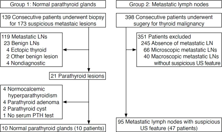
Methods: This retrospective study included 10 normal PTGs and 95 metastatic LNs from thyroid cancer showing suspicious US features. The echogenicity, echotexture, echogenic foci (calcifications), cystic change, abnormal vascularity, size, shape, and location were retrospectively assessed and compared between normal PTGs and metastatic LNs.
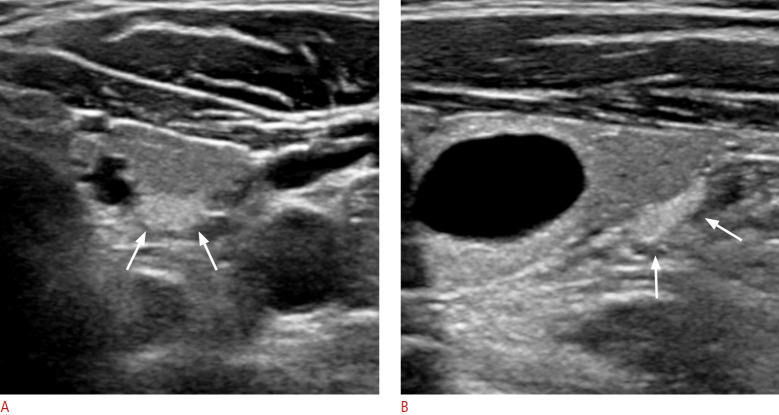
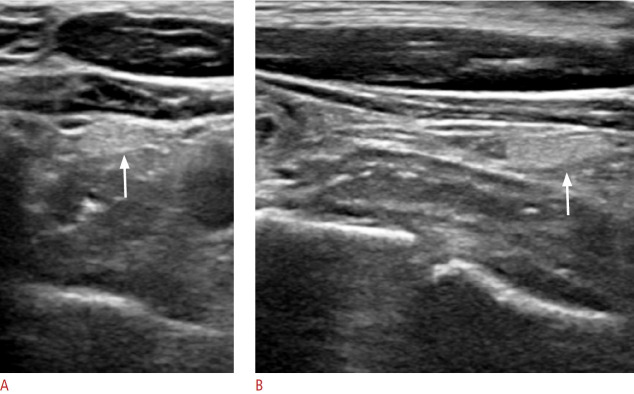
Results: The echogenicity of normal PTGs was significantly different from that of metastatic LNs (P<0.001). Normal PTGs exhibited marked hyperechogenicity (100%), homogeneous echotexture (80%), focal intraglandular hypoechogenicity (20%), ovoid shape (90%), and focal cystic change in one case (10%). The echogenicity of metastatic LNs was markedly hyperechoic (0%), moderately hyperechoic (15.8%), mildly hyperechoic (53.7%), and hypoechoic (28.4%). The size and long axis/short axis ratios of normal PTGs were significantly smaller and larger than those of metastatic LNs (P<0.01 and P=0.022, respectively).
Conclusion: Marked hyperechogenicity was found only in normal PTGs, and small, ovoid, markedly hyperechoic structures in the paramedian central neck characterized normal PTGs. Normal PTGs may be differentiated from metastatic LNs in thyroid cancer.
UltrasonographyMedicine-Radiology, Nuclear Medicine and Imaging
CiteScore
5.10
自引率
6.50%
发文量
78
审稿时长
15 weeks
期刊介绍:
Ultrasonography, the official English-language journal of the Korean Society of Ultrasound in Medicine (KSUM), is an international peer-reviewed academic journal dedicated to practice, research, technology, and education dealing with medical ultrasound. It is renamed from the Journal of Korean Society of Ultrasound in Medicine in January 2014, and published four times per year: January 1, April 1, July 1, and October 1. Original articles, technical notes, topical reviews, perspectives, pictorial essays, and timely editorial materials are published in Ultrasonography covering state-of-the-art content.
Ultrasonography aims to provide updated information on new diagnostic concepts and technical developments, including experimental animal studies using new equipment in addition to well-designed reviews of contemporary issues in patient care. Along with running KSUM Open, the annual international congress of KSUM, Ultrasonography also serves as a medium for cooperation among physicians and specialists from around the world who are focusing on various ultrasound technology and disease problems and relevant basic science.
 求助内容:
求助内容: 应助结果提醒方式:


