{"title":"可见光谱中热组织烧蚀的高对比度光谱光声表征。","authors":"Hyunjae Song, Tai-Kyong Song, Jeeun Kang","doi":"10.14366/usg.22171","DOIUrl":null,"url":null,"abstract":"<p><strong>Purpose: </strong>High-contrast tissue characterization of thermal ablation has been desired to evaluate therapeutic outcomes accurately. This paper presents a photoacoustic (PA) characterization of thermal tissue ablation in the visible spectrum, in which higher light absorbance can produce spectral contrast starker than in the near-infrared range.</p><p><strong>Methods: </strong>Ex vivo experiments were performed to measure visible PA spectra (480-700 nm) from fresh porcine liver tissues that received a thermal dose in a range of cumulative equivalent minutes at 43°C (CEM43). The local hemoglobin lobe area between 510-600 nm and wholespectral area under the curve were evaluated to represent the transition of hemoglobin into methemoglobin (MetHb) in the target tissue.</p><p><strong>Results: </strong>The thermal process below an estimated therapeutic CEM43 threshold (80-340 minutes) presented a progressive elevation of the PA spectrum and an eventual loss of local hemoglobin peaks in the visible spectrum, closer to the MetHb spectrum. Interestingly, an excessive CEM43 produced a substantial drop in the PA spectrum. In the spectral analysis, the visible spectrum yielded 13.9-34.1 times higher PA sensitivity and 1.42 times higher contrast change than at a near-infrared wavelength.</p><p><strong>Conclusion: </strong>This novel method of PA tissue characterization in the visible spectrum could be a potential modality to evaluate various thermal therapeutic modalities at high-contrast resolution.</p>","PeriodicalId":54227,"journal":{"name":"Ultrasonography","volume":"42 2","pages":"249-258"},"PeriodicalIF":2.5000,"publicationDate":"2023-04-01","publicationTypes":"Journal Article","fieldsOfStudy":null,"isOpenAccess":false,"openAccessPdf":"https://ftp.ncbi.nlm.nih.gov/pub/pmc/oa_pdf/59/92/usg-22171.PMC10071053.pdf","citationCount":"0","resultStr":"{\"title\":\"High-contrast spectroscopic photoacoustic characterization of thermal tissue ablation in the visible spectrum.\",\"authors\":\"Hyunjae Song, Tai-Kyong Song, Jeeun Kang\",\"doi\":\"10.14366/usg.22171\",\"DOIUrl\":null,\"url\":null,\"abstract\":\"<p><strong>Purpose: </strong>High-contrast tissue characterization of thermal ablation has been desired to evaluate therapeutic outcomes accurately. This paper presents a photoacoustic (PA) characterization of thermal tissue ablation in the visible spectrum, in which higher light absorbance can produce spectral contrast starker than in the near-infrared range.</p><p><strong>Methods: </strong>Ex vivo experiments were performed to measure visible PA spectra (480-700 nm) from fresh porcine liver tissues that received a thermal dose in a range of cumulative equivalent minutes at 43°C (CEM43). The local hemoglobin lobe area between 510-600 nm and wholespectral area under the curve were evaluated to represent the transition of hemoglobin into methemoglobin (MetHb) in the target tissue.</p><p><strong>Results: </strong>The thermal process below an estimated therapeutic CEM43 threshold (80-340 minutes) presented a progressive elevation of the PA spectrum and an eventual loss of local hemoglobin peaks in the visible spectrum, closer to the MetHb spectrum. Interestingly, an excessive CEM43 produced a substantial drop in the PA spectrum. In the spectral analysis, the visible spectrum yielded 13.9-34.1 times higher PA sensitivity and 1.42 times higher contrast change than at a near-infrared wavelength.</p><p><strong>Conclusion: </strong>This novel method of PA tissue characterization in the visible spectrum could be a potential modality to evaluate various thermal therapeutic modalities at high-contrast resolution.</p>\",\"PeriodicalId\":54227,\"journal\":{\"name\":\"Ultrasonography\",\"volume\":\"42 2\",\"pages\":\"249-258\"},\"PeriodicalIF\":2.5000,\"publicationDate\":\"2023-04-01\",\"publicationTypes\":\"Journal Article\",\"fieldsOfStudy\":null,\"isOpenAccess\":false,\"openAccessPdf\":\"https://ftp.ncbi.nlm.nih.gov/pub/pmc/oa_pdf/59/92/usg-22171.PMC10071053.pdf\",\"citationCount\":\"0\",\"resultStr\":null,\"platform\":\"Semanticscholar\",\"paperid\":null,\"PeriodicalName\":\"Ultrasonography\",\"FirstCategoryId\":\"3\",\"ListUrlMain\":\"https://doi.org/10.14366/usg.22171\",\"RegionNum\":3,\"RegionCategory\":\"医学\",\"ArticlePicture\":[],\"TitleCN\":null,\"AbstractTextCN\":null,\"PMCID\":null,\"EPubDate\":\"2022/11/29 0:00:00\",\"PubModel\":\"Epub\",\"JCR\":\"Q2\",\"JCRName\":\"RADIOLOGY, NUCLEAR MEDICINE & MEDICAL IMAGING\",\"Score\":null,\"Total\":0}","platform":"Semanticscholar","paperid":null,"PeriodicalName":"Ultrasonography","FirstCategoryId":"3","ListUrlMain":"https://doi.org/10.14366/usg.22171","RegionNum":3,"RegionCategory":"医学","ArticlePicture":[],"TitleCN":null,"AbstractTextCN":null,"PMCID":null,"EPubDate":"2022/11/29 0:00:00","PubModel":"Epub","JCR":"Q2","JCRName":"RADIOLOGY, NUCLEAR MEDICINE & MEDICAL IMAGING","Score":null,"Total":0}
引用次数: 0
摘要
目的:人们一直希望对热消融进行高对比度的组织表征,以准确评估治疗效果。本文介绍了可见光谱中热组织消融的光声(PA)表征,在可见光谱中,较高的光吸收率可产生比近红外范围更明显的光谱对比:活体实验测量了新鲜猪肝组织的可见 PA 光谱(480-700 nm),这些组织接受的热剂量范围为 43°C 下累积等效分钟(CEM43)。评估了 510-600 纳米之间的局部血红蛋白叶面积和曲线下的全谱面积,以表示目标组织中血红蛋白向高铁血红蛋白(MetHb)的转变:结果:在低于估计的治疗 CEM43 阈值(80-340 分钟)的热处理过程中,PA 光谱逐渐升高,最终在可见光谱中失去局部血红蛋白峰,更接近于高铁血红蛋白光谱。有趣的是,过量的 CEM43 会导致 PA 频谱大幅下降。在光谱分析中,与近红外波长相比,可见光谱的 PA 敏感度高 13.9-34.1 倍,对比度变化高 1.42 倍:结论:这种在可见光谱下描述 PA 组织特征的新方法可能是一种潜在的模式,可用于以高对比度分辨率评估各种热疗模式。
High-contrast spectroscopic photoacoustic characterization of thermal tissue ablation in the visible spectrum.
Purpose: High-contrast tissue characterization of thermal ablation has been desired to evaluate therapeutic outcomes accurately. This paper presents a photoacoustic (PA) characterization of thermal tissue ablation in the visible spectrum, in which higher light absorbance can produce spectral contrast starker than in the near-infrared range.
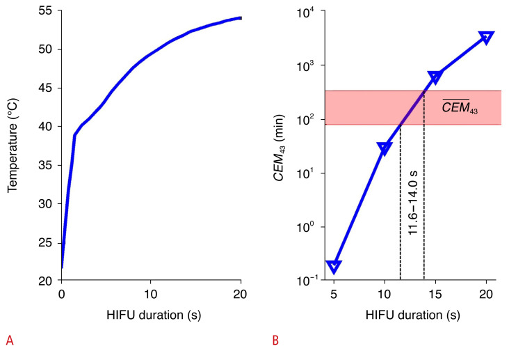
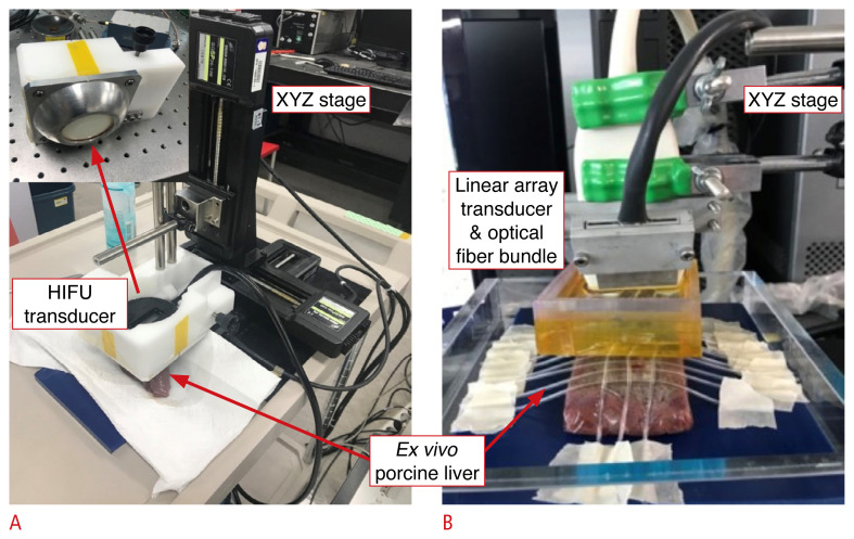
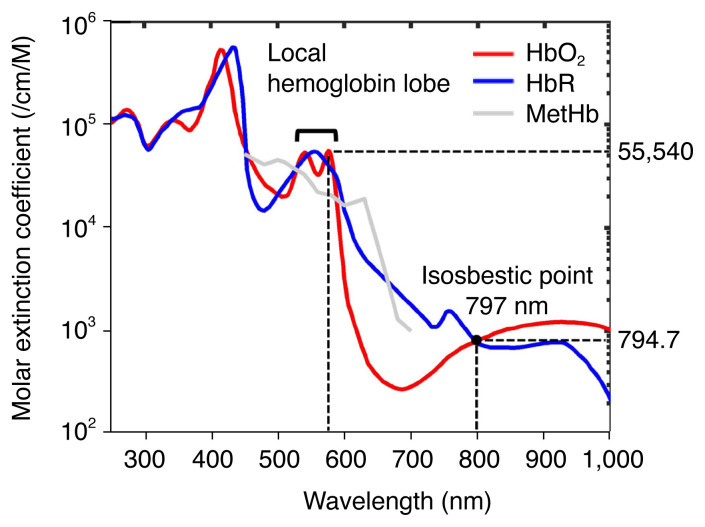
Methods: Ex vivo experiments were performed to measure visible PA spectra (480-700 nm) from fresh porcine liver tissues that received a thermal dose in a range of cumulative equivalent minutes at 43°C (CEM43). The local hemoglobin lobe area between 510-600 nm and wholespectral area under the curve were evaluated to represent the transition of hemoglobin into methemoglobin (MetHb) in the target tissue.
Results: The thermal process below an estimated therapeutic CEM43 threshold (80-340 minutes) presented a progressive elevation of the PA spectrum and an eventual loss of local hemoglobin peaks in the visible spectrum, closer to the MetHb spectrum. Interestingly, an excessive CEM43 produced a substantial drop in the PA spectrum. In the spectral analysis, the visible spectrum yielded 13.9-34.1 times higher PA sensitivity and 1.42 times higher contrast change than at a near-infrared wavelength.
Conclusion: This novel method of PA tissue characterization in the visible spectrum could be a potential modality to evaluate various thermal therapeutic modalities at high-contrast resolution.
UltrasonographyMedicine-Radiology, Nuclear Medicine and Imaging
CiteScore
5.10
自引率
6.50%
发文量
78
审稿时长
15 weeks
期刊介绍:
Ultrasonography, the official English-language journal of the Korean Society of Ultrasound in Medicine (KSUM), is an international peer-reviewed academic journal dedicated to practice, research, technology, and education dealing with medical ultrasound. It is renamed from the Journal of Korean Society of Ultrasound in Medicine in January 2014, and published four times per year: January 1, April 1, July 1, and October 1. Original articles, technical notes, topical reviews, perspectives, pictorial essays, and timely editorial materials are published in Ultrasonography covering state-of-the-art content.
Ultrasonography aims to provide updated information on new diagnostic concepts and technical developments, including experimental animal studies using new equipment in addition to well-designed reviews of contemporary issues in patient care. Along with running KSUM Open, the annual international congress of KSUM, Ultrasonography also serves as a medium for cooperation among physicians and specialists from around the world who are focusing on various ultrasound technology and disease problems and relevant basic science.
 求助内容:
求助内容: 应助结果提醒方式:


