Julian A Villalba, Adina Haramati, Michelle Garlin, Fabiola Reyes, Cameron D Wright, Abner Louissaint, Jeanne B Ackman
{"title":"切除胸腺囊肿的病灶内微出血在影像学上不确定。","authors":"Julian A Villalba, Adina Haramati, Michelle Garlin, Fabiola Reyes, Cameron D Wright, Abner Louissaint, Jeanne B Ackman","doi":"10.21037/med-22-42","DOIUrl":null,"url":null,"abstract":"<p><strong>Background: </strong>The propensity of thymic cysts to mimic solid thymic epithelial tumors (TETs) on computed tomography (CT), on account of attenuation values greater than water and thickened or calcified walls, can lead to non-therapeutic thymectomy. These lesions can fluctuate in volume, CT attenuation, and magnetic resonance imaging (MRI) signal over time. We hypothesized that spontaneous hemorrhage and resorption may contribute to their variable appearance over time.</p><p><strong>Methods: </strong>Completely excised thymic cysts were identified retrospectively over a 20-year period by their pathologic diagnosis. Cysts were excluded if they did not have available presurgical imaging, were not prevascular, were located within or contained an enhancing mass by imaging, or were of non-thymic origin upon microscopic review. Histopathological analysis of all available resected thymic cyst material and radiologic analysis of the cysts on pre-operative imaging were performed.</p><p><strong>Results: </strong>Upon application of exclusion criteria, we identified 18 thymic cysts from the initial 85 mediastinal cystic specimens. Most cysts were unilocular (11/15, 73%), showed turbid-to-semisolid, hemorrhagic fluid (10/12, 83%) and showed histopathological findings suggestive of intralesional microbleeding (14/18, 78%), remodeling (8/18, 44%), pathological wound healing/scarring of the capsule (16/18, 89%), and fat necrosis in the surrounding thymic tissue (12/18, 67%). On CT, 6/17 (35%) cysts demonstrated wall calcification. Sixty-five percent (11/17) had attenuation values ≥20 Hounsfield units (HU). Two of the 4 cysts imaged by MRI were T1-isointense, one was mixed hyper- and isointense, and one T1-hypointense to muscle, with iso- and hyperintensity indicating hemorrhagic or proteinaceous content. Twenty-five percent (1/4) of cyst walls imaged by MRI were T1/T2-hypointense, indicating presence of calcification, hemosiderin, and/or fibrosis.</p><p><strong>Conclusions: </strong>Resected thymic cysts in this cohort often showed features suggestive of intralesional microbleeding, inflammation, and fibrosis, which may explain their appearance and behavior over time on CT and MRI.</p>","PeriodicalId":74139,"journal":{"name":"Mediastinum (Hong Kong, China)","volume":"7 ","pages":"13"},"PeriodicalIF":0.0000,"publicationDate":"2023-01-01","publicationTypes":"Journal Article","fieldsOfStudy":null,"isOpenAccess":false,"openAccessPdf":"https://ftp.ncbi.nlm.nih.gov/pub/pmc/oa_pdf/01/fd/med-07-13.PMC10226889.pdf","citationCount":"0","resultStr":"{\"title\":\"Intralesional microbleeding in resected thymic cysts indeterminate on imaging.\",\"authors\":\"Julian A Villalba, Adina Haramati, Michelle Garlin, Fabiola Reyes, Cameron D Wright, Abner Louissaint, Jeanne B Ackman\",\"doi\":\"10.21037/med-22-42\",\"DOIUrl\":null,\"url\":null,\"abstract\":\"<p><strong>Background: </strong>The propensity of thymic cysts to mimic solid thymic epithelial tumors (TETs) on computed tomography (CT), on account of attenuation values greater than water and thickened or calcified walls, can lead to non-therapeutic thymectomy. These lesions can fluctuate in volume, CT attenuation, and magnetic resonance imaging (MRI) signal over time. We hypothesized that spontaneous hemorrhage and resorption may contribute to their variable appearance over time.</p><p><strong>Methods: </strong>Completely excised thymic cysts were identified retrospectively over a 20-year period by their pathologic diagnosis. Cysts were excluded if they did not have available presurgical imaging, were not prevascular, were located within or contained an enhancing mass by imaging, or were of non-thymic origin upon microscopic review. Histopathological analysis of all available resected thymic cyst material and radiologic analysis of the cysts on pre-operative imaging were performed.</p><p><strong>Results: </strong>Upon application of exclusion criteria, we identified 18 thymic cysts from the initial 85 mediastinal cystic specimens. Most cysts were unilocular (11/15, 73%), showed turbid-to-semisolid, hemorrhagic fluid (10/12, 83%) and showed histopathological findings suggestive of intralesional microbleeding (14/18, 78%), remodeling (8/18, 44%), pathological wound healing/scarring of the capsule (16/18, 89%), and fat necrosis in the surrounding thymic tissue (12/18, 67%). On CT, 6/17 (35%) cysts demonstrated wall calcification. Sixty-five percent (11/17) had attenuation values ≥20 Hounsfield units (HU). Two of the 4 cysts imaged by MRI were T1-isointense, one was mixed hyper- and isointense, and one T1-hypointense to muscle, with iso- and hyperintensity indicating hemorrhagic or proteinaceous content. Twenty-five percent (1/4) of cyst walls imaged by MRI were T1/T2-hypointense, indicating presence of calcification, hemosiderin, and/or fibrosis.</p><p><strong>Conclusions: </strong>Resected thymic cysts in this cohort often showed features suggestive of intralesional microbleeding, inflammation, and fibrosis, which may explain their appearance and behavior over time on CT and MRI.</p>\",\"PeriodicalId\":74139,\"journal\":{\"name\":\"Mediastinum (Hong Kong, China)\",\"volume\":\"7 \",\"pages\":\"13\"},\"PeriodicalIF\":0.0000,\"publicationDate\":\"2023-01-01\",\"publicationTypes\":\"Journal Article\",\"fieldsOfStudy\":null,\"isOpenAccess\":false,\"openAccessPdf\":\"https://ftp.ncbi.nlm.nih.gov/pub/pmc/oa_pdf/01/fd/med-07-13.PMC10226889.pdf\",\"citationCount\":\"0\",\"resultStr\":null,\"platform\":\"Semanticscholar\",\"paperid\":null,\"PeriodicalName\":\"Mediastinum (Hong Kong, China)\",\"FirstCategoryId\":\"1085\",\"ListUrlMain\":\"https://doi.org/10.21037/med-22-42\",\"RegionNum\":0,\"RegionCategory\":null,\"ArticlePicture\":[],\"TitleCN\":null,\"AbstractTextCN\":null,\"PMCID\":null,\"EPubDate\":\"\",\"PubModel\":\"\",\"JCR\":\"\",\"JCRName\":\"\",\"Score\":null,\"Total\":0}","platform":"Semanticscholar","paperid":null,"PeriodicalName":"Mediastinum (Hong Kong, China)","FirstCategoryId":"1085","ListUrlMain":"https://doi.org/10.21037/med-22-42","RegionNum":0,"RegionCategory":null,"ArticlePicture":[],"TitleCN":null,"AbstractTextCN":null,"PMCID":null,"EPubDate":"","PubModel":"","JCR":"","JCRName":"","Score":null,"Total":0}
Intralesional microbleeding in resected thymic cysts indeterminate on imaging.
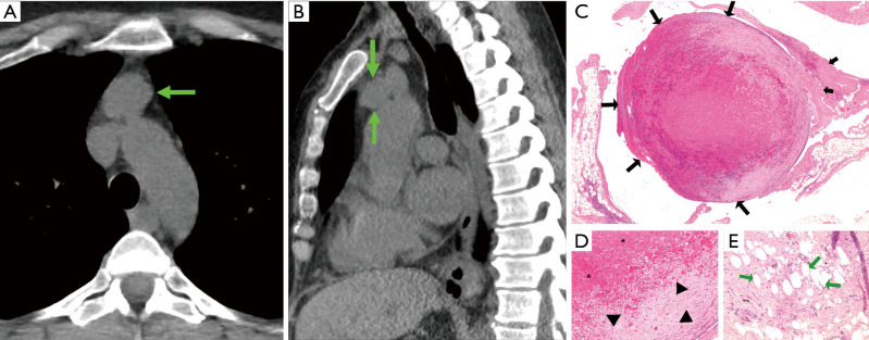
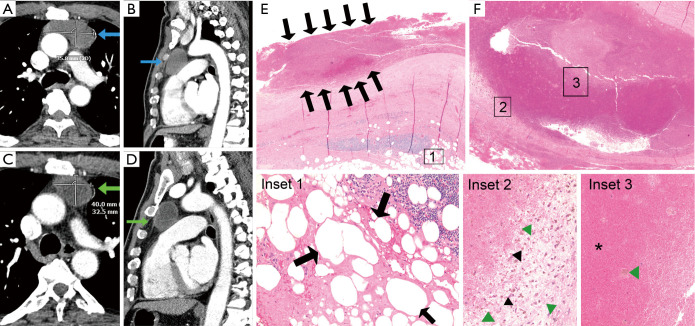
Background: The propensity of thymic cysts to mimic solid thymic epithelial tumors (TETs) on computed tomography (CT), on account of attenuation values greater than water and thickened or calcified walls, can lead to non-therapeutic thymectomy. These lesions can fluctuate in volume, CT attenuation, and magnetic resonance imaging (MRI) signal over time. We hypothesized that spontaneous hemorrhage and resorption may contribute to their variable appearance over time.
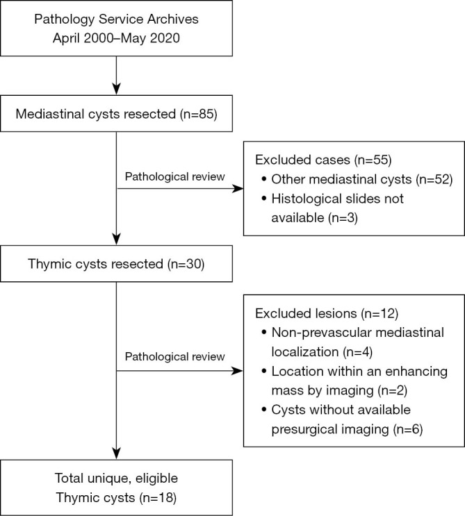
Methods: Completely excised thymic cysts were identified retrospectively over a 20-year period by their pathologic diagnosis. Cysts were excluded if they did not have available presurgical imaging, were not prevascular, were located within or contained an enhancing mass by imaging, or were of non-thymic origin upon microscopic review. Histopathological analysis of all available resected thymic cyst material and radiologic analysis of the cysts on pre-operative imaging were performed.
Results: Upon application of exclusion criteria, we identified 18 thymic cysts from the initial 85 mediastinal cystic specimens. Most cysts were unilocular (11/15, 73%), showed turbid-to-semisolid, hemorrhagic fluid (10/12, 83%) and showed histopathological findings suggestive of intralesional microbleeding (14/18, 78%), remodeling (8/18, 44%), pathological wound healing/scarring of the capsule (16/18, 89%), and fat necrosis in the surrounding thymic tissue (12/18, 67%). On CT, 6/17 (35%) cysts demonstrated wall calcification. Sixty-five percent (11/17) had attenuation values ≥20 Hounsfield units (HU). Two of the 4 cysts imaged by MRI were T1-isointense, one was mixed hyper- and isointense, and one T1-hypointense to muscle, with iso- and hyperintensity indicating hemorrhagic or proteinaceous content. Twenty-five percent (1/4) of cyst walls imaged by MRI were T1/T2-hypointense, indicating presence of calcification, hemosiderin, and/or fibrosis.
Conclusions: Resected thymic cysts in this cohort often showed features suggestive of intralesional microbleeding, inflammation, and fibrosis, which may explain their appearance and behavior over time on CT and MRI.

 求助内容:
求助内容: 应助结果提醒方式:


