Jacob Sjölin, Marianne Jonsson, Charlotta Orback, Anders Oldfors, Anders Jeppsson, Jane Synnergren, Victoria Rotter Sopasakis, Kristina Vukusic
{"title":"干细胞生态位相关生物标志物在人三尖瓣基部的表达。","authors":"Jacob Sjölin, Marianne Jonsson, Charlotta Orback, Anders Oldfors, Anders Jeppsson, Jane Synnergren, Victoria Rotter Sopasakis, Kristina Vukusic","doi":"10.1089/scd.2022.0253","DOIUrl":null,"url":null,"abstract":"<p><p>Stem cell niches have been thoroughly investigated in tissue with high regenerative capacity but not in tissues where cell turnover is slow, such as the human heart. The left AtrioVentricular junction (AVj), the base of the mitral valve, has previously been proposed as a niche region for cardiac progenitors in the adult human heart. In the present study, we explore the right side of the human heart, the base of the tricuspid valve, to investigate the potential of this region as a progenitor niche. Paired biopsies from explanted human hearts were collected from multi-organ donors (<i>N</i> = 12). The lateral side of the AVj, right atria (RA), and right ventricle (RV) were compared for the expression of stem cell niche-related biomarkers using RNA sequencing. Gene expression data indicated upregulation of genes related to embryonic development and extracellular matrix (ECM) composition in the proposed niche region, that is, the AVj. In addition, immunohistochemistry showed high expression of the fetal cardiac markers MDR1, SSEA4, and WT1 within the same region. Nuclear expression of HIF1α was detected suggesting hypoxia. Rare cells were found with the co-staining of the proliferation marker PCNA and Ki67 with cardiomyocyte nuclei marker PCM1 and cardiac Troponin T (cTnT), indicating proliferation of small cardiomyocytes. WT1+/cTnT+ and SSEA4+/cTnT+ cells were also found, suggesting cardiomyocyte-specific progenitors. The expression of the stem cell markers gradually decreased with distance from the tricuspid valve. No expression of these markers was observed in the RV tissue. In summary, the base of the tricuspid valve is an ECM-rich region containing cells with expression of several stem cell niche-associated markers. Co-expression of stem cell markers with cTnT indicates cardiomyocyte-specific progenitors. We previously reported similar data from the base of the mitral valve and thus propose that human adult cardiomyocyte progenitors reside around both atrioventricular valves.</p>","PeriodicalId":21934,"journal":{"name":"Stem cells and development","volume":"32 5-6","pages":"140-151"},"PeriodicalIF":2.0000,"publicationDate":"2023-03-01","publicationTypes":"Journal Article","fieldsOfStudy":null,"isOpenAccess":false,"openAccessPdf":"https://www.ncbi.nlm.nih.gov/pmc/articles/PMC9986114/pdf/","citationCount":"0","resultStr":"{\"title\":\"Expression of Stem Cell Niche-Related Biomarkers at the Base of the Human Tricuspid Valve.\",\"authors\":\"Jacob Sjölin, Marianne Jonsson, Charlotta Orback, Anders Oldfors, Anders Jeppsson, Jane Synnergren, Victoria Rotter Sopasakis, Kristina Vukusic\",\"doi\":\"10.1089/scd.2022.0253\",\"DOIUrl\":null,\"url\":null,\"abstract\":\"<p><p>Stem cell niches have been thoroughly investigated in tissue with high regenerative capacity but not in tissues where cell turnover is slow, such as the human heart. The left AtrioVentricular junction (AVj), the base of the mitral valve, has previously been proposed as a niche region for cardiac progenitors in the adult human heart. In the present study, we explore the right side of the human heart, the base of the tricuspid valve, to investigate the potential of this region as a progenitor niche. Paired biopsies from explanted human hearts were collected from multi-organ donors (<i>N</i> = 12). The lateral side of the AVj, right atria (RA), and right ventricle (RV) were compared for the expression of stem cell niche-related biomarkers using RNA sequencing. Gene expression data indicated upregulation of genes related to embryonic development and extracellular matrix (ECM) composition in the proposed niche region, that is, the AVj. In addition, immunohistochemistry showed high expression of the fetal cardiac markers MDR1, SSEA4, and WT1 within the same region. Nuclear expression of HIF1α was detected suggesting hypoxia. Rare cells were found with the co-staining of the proliferation marker PCNA and Ki67 with cardiomyocyte nuclei marker PCM1 and cardiac Troponin T (cTnT), indicating proliferation of small cardiomyocytes. WT1+/cTnT+ and SSEA4+/cTnT+ cells were also found, suggesting cardiomyocyte-specific progenitors. The expression of the stem cell markers gradually decreased with distance from the tricuspid valve. No expression of these markers was observed in the RV tissue. In summary, the base of the tricuspid valve is an ECM-rich region containing cells with expression of several stem cell niche-associated markers. Co-expression of stem cell markers with cTnT indicates cardiomyocyte-specific progenitors. We previously reported similar data from the base of the mitral valve and thus propose that human adult cardiomyocyte progenitors reside around both atrioventricular valves.</p>\",\"PeriodicalId\":21934,\"journal\":{\"name\":\"Stem cells and development\",\"volume\":\"32 5-6\",\"pages\":\"140-151\"},\"PeriodicalIF\":2.0000,\"publicationDate\":\"2023-03-01\",\"publicationTypes\":\"Journal Article\",\"fieldsOfStudy\":null,\"isOpenAccess\":false,\"openAccessPdf\":\"https://www.ncbi.nlm.nih.gov/pmc/articles/PMC9986114/pdf/\",\"citationCount\":\"0\",\"resultStr\":null,\"platform\":\"Semanticscholar\",\"paperid\":null,\"PeriodicalName\":\"Stem cells and development\",\"FirstCategoryId\":\"3\",\"ListUrlMain\":\"https://doi.org/10.1089/scd.2022.0253\",\"RegionNum\":3,\"RegionCategory\":\"医学\",\"ArticlePicture\":[],\"TitleCN\":null,\"AbstractTextCN\":null,\"PMCID\":null,\"EPubDate\":\"\",\"PubModel\":\"\",\"JCR\":\"Q3\",\"JCRName\":\"CELL & TISSUE ENGINEERING\",\"Score\":null,\"Total\":0}","platform":"Semanticscholar","paperid":null,"PeriodicalName":"Stem cells and development","FirstCategoryId":"3","ListUrlMain":"https://doi.org/10.1089/scd.2022.0253","RegionNum":3,"RegionCategory":"医学","ArticlePicture":[],"TitleCN":null,"AbstractTextCN":null,"PMCID":null,"EPubDate":"","PubModel":"","JCR":"Q3","JCRName":"CELL & TISSUE ENGINEERING","Score":null,"Total":0}
Expression of Stem Cell Niche-Related Biomarkers at the Base of the Human Tricuspid Valve.
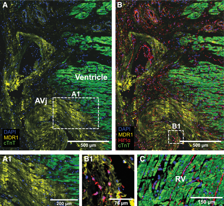
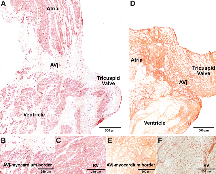
Stem cell niches have been thoroughly investigated in tissue with high regenerative capacity but not in tissues where cell turnover is slow, such as the human heart. The left AtrioVentricular junction (AVj), the base of the mitral valve, has previously been proposed as a niche region for cardiac progenitors in the adult human heart. In the present study, we explore the right side of the human heart, the base of the tricuspid valve, to investigate the potential of this region as a progenitor niche. Paired biopsies from explanted human hearts were collected from multi-organ donors (N = 12). The lateral side of the AVj, right atria (RA), and right ventricle (RV) were compared for the expression of stem cell niche-related biomarkers using RNA sequencing. Gene expression data indicated upregulation of genes related to embryonic development and extracellular matrix (ECM) composition in the proposed niche region, that is, the AVj. In addition, immunohistochemistry showed high expression of the fetal cardiac markers MDR1, SSEA4, and WT1 within the same region. Nuclear expression of HIF1α was detected suggesting hypoxia. Rare cells were found with the co-staining of the proliferation marker PCNA and Ki67 with cardiomyocyte nuclei marker PCM1 and cardiac Troponin T (cTnT), indicating proliferation of small cardiomyocytes. WT1+/cTnT+ and SSEA4+/cTnT+ cells were also found, suggesting cardiomyocyte-specific progenitors. The expression of the stem cell markers gradually decreased with distance from the tricuspid valve. No expression of these markers was observed in the RV tissue. In summary, the base of the tricuspid valve is an ECM-rich region containing cells with expression of several stem cell niche-associated markers. Co-expression of stem cell markers with cTnT indicates cardiomyocyte-specific progenitors. We previously reported similar data from the base of the mitral valve and thus propose that human adult cardiomyocyte progenitors reside around both atrioventricular valves.
期刊介绍:
Stem Cells and Development is globally recognized as the trusted source for critical, even controversial coverage of emerging hypotheses and novel findings. With a focus on stem cells of all tissue types and their potential therapeutic applications, the Journal provides clinical, basic, and translational scientists with cutting-edge research and findings.
Stem Cells and Development coverage includes:
Embryogenesis and adult counterparts of this process
Physical processes linking stem cells, primary cell function, and structural development
Hypotheses exploring the relationship between genotype and phenotype
Development of vasculature, CNS, and other germ layer development and defects
Pluripotentiality of embryonic and somatic stem cells
The role of genetic and epigenetic factors in development

 求助内容:
求助内容: 应助结果提醒方式:


