Yusuke S Hori, John S Albanese, John G Meara, Mark R Proctor
{"title":"小儿神经外科影像:鼻额交界处隐匿性骨内皮样囊肿。","authors":"Yusuke S Hori, John S Albanese, John G Meara, Mark R Proctor","doi":"10.1159/000528440","DOIUrl":null,"url":null,"abstract":"INTRODUCTION\nAn extension of the dermoid cyst below the nasal bone has been identified in 10% of patients in a large series of nasal dermoid cysts, but these are generally easily identifiable and connected to the tract. To date, no previous reports have documented a case with intraosseous dermoid cyst which was completely hidden in the nasal bone.\n\n\nCASE PRESENTATION\nAn 8 year-old previously healthy female was initially found to have a small pit on her nasal dorsum. The lesion developed local infection and she was initially treated with antibiotics two years prior to the current presentation. The lesion was diagnosed as a dermal sinus tract, and surgical removal was conducted at an outside hospital. In retrospect the pre-operative work-up imaging showed an occult intraosseous nasal bone extension, however, this was not appreciated at the initial surgery. She experienced repeat infections and underwent a second surgery with exploration under the nasal bones, however, the patient experienced recurrent postoperative local infections. The family presented to our institution for a second opinion. Our images interestingly illustrate the nasal dermoid cyst extending into the nasal bone at the nasofrontal junction without detectable extraosseous sub-nasal bone extension on the imaging. The patient proceeded with a third surgery for complete removal of the lesion via an extended vertical nasal incision. The nasal bones were removed in their entirety, the occult dermoid cyst with a small tract was located in the nasal bones and the undersurface of the bones were completely debrided. No intracranial extension was observed after careful investigation of the skull base. Particulate corticocancellous bone was used with fibrin sealant to reconstruct the defect. The nasal bones were then replaced. The pathology results were consistent with a dermoid cyst. The post-operative course was uncomplicated and she has not had a recurrence after the third surgery.\n\n\nDISCUSSION/CONCLUSION\nThis case, despite an exploration under the nasal bones, initially received incomplete resection and experienced multiple infections because of failure to appreciate the portion hidden in the nasal bones. Our case was successfully treated with ostectomy of nasal bones without recurrence and complications. This procedure allows unobstructed visualization of the entire cyst leading to the complete removal of the lesion. This is an instructive case to show that portions of the cyst may remain hidden and lead to recurrent infection, and complete resection with sufficient exposure of the entire lesion is needed to successfully treat this condition.","PeriodicalId":54631,"journal":{"name":"Pediatric Neurosurgery","volume":"58 1","pages":"58-60"},"PeriodicalIF":0.9000,"publicationDate":"2023-01-01","publicationTypes":"Journal Article","fieldsOfStudy":null,"isOpenAccess":false,"openAccessPdf":"https://www.ncbi.nlm.nih.gov/pmc/articles/PMC10064384/pdf/","citationCount":"0","resultStr":"{\"title\":\"Images in Pediatric Neurosurgery: Occult Intraosseous Dermoid Cyst at the Nasofrontal Junction.\",\"authors\":\"Yusuke S Hori, John S Albanese, John G Meara, Mark R Proctor\",\"doi\":\"10.1159/000528440\",\"DOIUrl\":null,\"url\":null,\"abstract\":\"INTRODUCTION\\nAn extension of the dermoid cyst below the nasal bone has been identified in 10% of patients in a large series of nasal dermoid cysts, but these are generally easily identifiable and connected to the tract. To date, no previous reports have documented a case with intraosseous dermoid cyst which was completely hidden in the nasal bone.\\n\\n\\nCASE PRESENTATION\\nAn 8 year-old previously healthy female was initially found to have a small pit on her nasal dorsum. The lesion developed local infection and she was initially treated with antibiotics two years prior to the current presentation. The lesion was diagnosed as a dermal sinus tract, and surgical removal was conducted at an outside hospital. In retrospect the pre-operative work-up imaging showed an occult intraosseous nasal bone extension, however, this was not appreciated at the initial surgery. She experienced repeat infections and underwent a second surgery with exploration under the nasal bones, however, the patient experienced recurrent postoperative local infections. The family presented to our institution for a second opinion. Our images interestingly illustrate the nasal dermoid cyst extending into the nasal bone at the nasofrontal junction without detectable extraosseous sub-nasal bone extension on the imaging. The patient proceeded with a third surgery for complete removal of the lesion via an extended vertical nasal incision. The nasal bones were removed in their entirety, the occult dermoid cyst with a small tract was located in the nasal bones and the undersurface of the bones were completely debrided. No intracranial extension was observed after careful investigation of the skull base. Particulate corticocancellous bone was used with fibrin sealant to reconstruct the defect. The nasal bones were then replaced. The pathology results were consistent with a dermoid cyst. The post-operative course was uncomplicated and she has not had a recurrence after the third surgery.\\n\\n\\nDISCUSSION/CONCLUSION\\nThis case, despite an exploration under the nasal bones, initially received incomplete resection and experienced multiple infections because of failure to appreciate the portion hidden in the nasal bones. Our case was successfully treated with ostectomy of nasal bones without recurrence and complications. This procedure allows unobstructed visualization of the entire cyst leading to the complete removal of the lesion. This is an instructive case to show that portions of the cyst may remain hidden and lead to recurrent infection, and complete resection with sufficient exposure of the entire lesion is needed to successfully treat this condition.\",\"PeriodicalId\":54631,\"journal\":{\"name\":\"Pediatric Neurosurgery\",\"volume\":\"58 1\",\"pages\":\"58-60\"},\"PeriodicalIF\":0.9000,\"publicationDate\":\"2023-01-01\",\"publicationTypes\":\"Journal Article\",\"fieldsOfStudy\":null,\"isOpenAccess\":false,\"openAccessPdf\":\"https://www.ncbi.nlm.nih.gov/pmc/articles/PMC10064384/pdf/\",\"citationCount\":\"0\",\"resultStr\":null,\"platform\":\"Semanticscholar\",\"paperid\":null,\"PeriodicalName\":\"Pediatric Neurosurgery\",\"FirstCategoryId\":\"3\",\"ListUrlMain\":\"https://doi.org/10.1159/000528440\",\"RegionNum\":4,\"RegionCategory\":\"医学\",\"ArticlePicture\":[],\"TitleCN\":null,\"AbstractTextCN\":null,\"PMCID\":null,\"EPubDate\":\"\",\"PubModel\":\"\",\"JCR\":\"Q4\",\"JCRName\":\"CLINICAL NEUROLOGY\",\"Score\":null,\"Total\":0}","platform":"Semanticscholar","paperid":null,"PeriodicalName":"Pediatric Neurosurgery","FirstCategoryId":"3","ListUrlMain":"https://doi.org/10.1159/000528440","RegionNum":4,"RegionCategory":"医学","ArticlePicture":[],"TitleCN":null,"AbstractTextCN":null,"PMCID":null,"EPubDate":"","PubModel":"","JCR":"Q4","JCRName":"CLINICAL NEUROLOGY","Score":null,"Total":0}
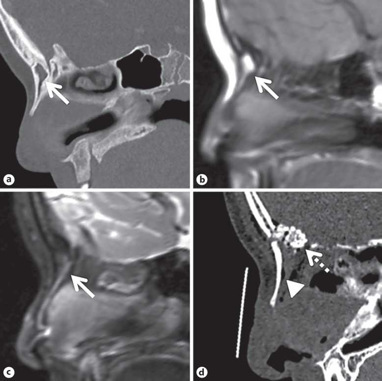
Images in Pediatric Neurosurgery: Occult Intraosseous Dermoid Cyst at the Nasofrontal Junction.
INTRODUCTION
An extension of the dermoid cyst below the nasal bone has been identified in 10% of patients in a large series of nasal dermoid cysts, but these are generally easily identifiable and connected to the tract. To date, no previous reports have documented a case with intraosseous dermoid cyst which was completely hidden in the nasal bone.
CASE PRESENTATION
An 8 year-old previously healthy female was initially found to have a small pit on her nasal dorsum. The lesion developed local infection and she was initially treated with antibiotics two years prior to the current presentation. The lesion was diagnosed as a dermal sinus tract, and surgical removal was conducted at an outside hospital. In retrospect the pre-operative work-up imaging showed an occult intraosseous nasal bone extension, however, this was not appreciated at the initial surgery. She experienced repeat infections and underwent a second surgery with exploration under the nasal bones, however, the patient experienced recurrent postoperative local infections. The family presented to our institution for a second opinion. Our images interestingly illustrate the nasal dermoid cyst extending into the nasal bone at the nasofrontal junction without detectable extraosseous sub-nasal bone extension on the imaging. The patient proceeded with a third surgery for complete removal of the lesion via an extended vertical nasal incision. The nasal bones were removed in their entirety, the occult dermoid cyst with a small tract was located in the nasal bones and the undersurface of the bones were completely debrided. No intracranial extension was observed after careful investigation of the skull base. Particulate corticocancellous bone was used with fibrin sealant to reconstruct the defect. The nasal bones were then replaced. The pathology results were consistent with a dermoid cyst. The post-operative course was uncomplicated and she has not had a recurrence after the third surgery.
DISCUSSION/CONCLUSION
This case, despite an exploration under the nasal bones, initially received incomplete resection and experienced multiple infections because of failure to appreciate the portion hidden in the nasal bones. Our case was successfully treated with ostectomy of nasal bones without recurrence and complications. This procedure allows unobstructed visualization of the entire cyst leading to the complete removal of the lesion. This is an instructive case to show that portions of the cyst may remain hidden and lead to recurrent infection, and complete resection with sufficient exposure of the entire lesion is needed to successfully treat this condition.
期刊介绍:
Articles in ''Pediatric Neurosurgery'' strives to publish new information and observations in pediatric neurosurgery and the allied fields of neurology, neuroradiology and neuropathology as they relate to the etiology of neurologic diseases and the operative care of affected patients. In addition to experimental and clinical studies, the journal presents critical reviews which provide the reader with an update on selected topics as well as case histories and reports on advances in methodology and technique. This thought-provoking focus encourages dissemination of information from neurosurgeons and neuroscientists around the world that will be of interest to clinicians and researchers concerned with pediatric, congenital, and developmental diseases of the nervous system.
 求助内容:
求助内容: 应助结果提醒方式:


