Nikolaos Galanakis, Nikolaos Kontopodis, Nikolas Matthaiou, Dimitrios Tsetis, Elias Kehagias
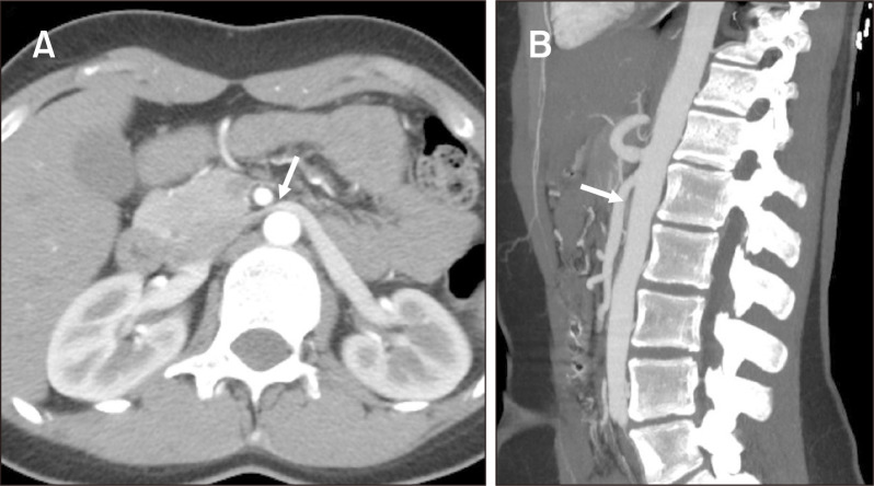
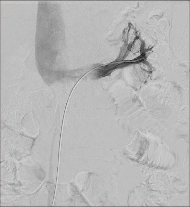
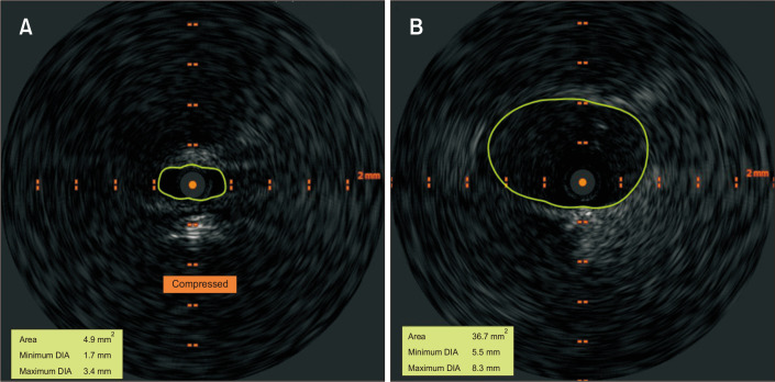
{"title":"一位年轻患者胡桃夹子综合征的多模态评估。","authors":"Nikolaos Galanakis, Nikolaos Kontopodis, Nikolas Matthaiou, Dimitrios Tsetis, Elias Kehagias","doi":"10.5758/vsi.230034","DOIUrl":null,"url":null,"abstract":"A 17-year-old female presented to University Hospital of Heraklion with symptoms of intermittent hematuria and moderate left lumbar pain. She denied a history of trauma and hematological and kidney diseases. She received no regular medications. Laboratory examinations were normal, and the patient underwent computed tomographic angiography (CTA). CTA demonstrated left renal vein (LRV) stenosis between the superior mesenteric artery (SMA) and aorta and post-stenotic LRV dilatation (Fig. 1). Moreover, a significantly reduced aortic-SMA angle was found (15°-20°). These findings suggest the presence of nutcracker syndrome (NCS). However, because of the patient’s young age and possible complications of surgical treatment, further evaluation with selective digital subtraction angiography (DSA) of the LRV was performed through percutaneous right common femoral vein access. However, it did not reveal any significant LRV stenosis (Fig. 2). Intravascular ultrasound (IVUS, Visions PV.035; Philips Volcano) was performed and demonstrated severe luminal narrowing in the proximal LRV (Fig. 3). Moreover, a significant pressure gradient (approximately 6 mmHg) between LRV and inferior Im ge of Vacular Srgery Multimodality Assessment of Nutcracker Syndrome in a Young Patient","PeriodicalId":52311,"journal":{"name":"Vascular Specialist International","volume":"39 ","pages":"11"},"PeriodicalIF":0.8000,"publicationDate":"2023-05-30","publicationTypes":"Journal Article","fieldsOfStudy":null,"isOpenAccess":false,"openAccessPdf":"https://ftp.ncbi.nlm.nih.gov/pub/pmc/oa_pdf/37/c3/vsi-39-11.PMC10227710.pdf","citationCount":"0","resultStr":"{\"title\":\"Multimodality Assessment of Nutcracker Syndrome in a Young Patient.\",\"authors\":\"Nikolaos Galanakis, Nikolaos Kontopodis, Nikolas Matthaiou, Dimitrios Tsetis, Elias Kehagias\",\"doi\":\"10.5758/vsi.230034\",\"DOIUrl\":null,\"url\":null,\"abstract\":\"A 17-year-old female presented to University Hospital of Heraklion with symptoms of intermittent hematuria and moderate left lumbar pain. She denied a history of trauma and hematological and kidney diseases. She received no regular medications. Laboratory examinations were normal, and the patient underwent computed tomographic angiography (CTA). CTA demonstrated left renal vein (LRV) stenosis between the superior mesenteric artery (SMA) and aorta and post-stenotic LRV dilatation (Fig. 1). Moreover, a significantly reduced aortic-SMA angle was found (15°-20°). These findings suggest the presence of nutcracker syndrome (NCS). However, because of the patient’s young age and possible complications of surgical treatment, further evaluation with selective digital subtraction angiography (DSA) of the LRV was performed through percutaneous right common femoral vein access. However, it did not reveal any significant LRV stenosis (Fig. 2). Intravascular ultrasound (IVUS, Visions PV.035; Philips Volcano) was performed and demonstrated severe luminal narrowing in the proximal LRV (Fig. 3). Moreover, a significant pressure gradient (approximately 6 mmHg) between LRV and inferior Im ge of Vacular Srgery Multimodality Assessment of Nutcracker Syndrome in a Young Patient\",\"PeriodicalId\":52311,\"journal\":{\"name\":\"Vascular Specialist International\",\"volume\":\"39 \",\"pages\":\"11\"},\"PeriodicalIF\":0.8000,\"publicationDate\":\"2023-05-30\",\"publicationTypes\":\"Journal Article\",\"fieldsOfStudy\":null,\"isOpenAccess\":false,\"openAccessPdf\":\"https://ftp.ncbi.nlm.nih.gov/pub/pmc/oa_pdf/37/c3/vsi-39-11.PMC10227710.pdf\",\"citationCount\":\"0\",\"resultStr\":null,\"platform\":\"Semanticscholar\",\"paperid\":null,\"PeriodicalName\":\"Vascular Specialist International\",\"FirstCategoryId\":\"1085\",\"ListUrlMain\":\"https://doi.org/10.5758/vsi.230034\",\"RegionNum\":0,\"RegionCategory\":null,\"ArticlePicture\":[],\"TitleCN\":null,\"AbstractTextCN\":null,\"PMCID\":null,\"EPubDate\":\"\",\"PubModel\":\"\",\"JCR\":\"Q4\",\"JCRName\":\"PERIPHERAL VASCULAR DISEASE\",\"Score\":null,\"Total\":0}","platform":"Semanticscholar","paperid":null,"PeriodicalName":"Vascular Specialist International","FirstCategoryId":"1085","ListUrlMain":"https://doi.org/10.5758/vsi.230034","RegionNum":0,"RegionCategory":null,"ArticlePicture":[],"TitleCN":null,"AbstractTextCN":null,"PMCID":null,"EPubDate":"","PubModel":"","JCR":"Q4","JCRName":"PERIPHERAL VASCULAR DISEASE","Score":null,"Total":0}
Multimodality Assessment of Nutcracker Syndrome in a Young Patient.
A 17-year-old female presented to University Hospital of Heraklion with symptoms of intermittent hematuria and moderate left lumbar pain. She denied a history of trauma and hematological and kidney diseases. She received no regular medications. Laboratory examinations were normal, and the patient underwent computed tomographic angiography (CTA). CTA demonstrated left renal vein (LRV) stenosis between the superior mesenteric artery (SMA) and aorta and post-stenotic LRV dilatation (Fig. 1). Moreover, a significantly reduced aortic-SMA angle was found (15°-20°). These findings suggest the presence of nutcracker syndrome (NCS). However, because of the patient’s young age and possible complications of surgical treatment, further evaluation with selective digital subtraction angiography (DSA) of the LRV was performed through percutaneous right common femoral vein access. However, it did not reveal any significant LRV stenosis (Fig. 2). Intravascular ultrasound (IVUS, Visions PV.035; Philips Volcano) was performed and demonstrated severe luminal narrowing in the proximal LRV (Fig. 3). Moreover, a significant pressure gradient (approximately 6 mmHg) between LRV and inferior Im ge of Vacular Srgery Multimodality Assessment of Nutcracker Syndrome in a Young Patient