Kevin K Kumar, Angus Toland, Nancy Fischbein, Martha Morrell, Jeremy J Heit, Donald E Born, Gary K Steinberg
{"title":"与脑裂畸形相关的血管异常、脂肪瘤和多微脑回畸形:发育和诊断见解。说明性案例。","authors":"Kevin K Kumar, Angus Toland, Nancy Fischbein, Martha Morrell, Jeremy J Heit, Donald E Born, Gary K Steinberg","doi":"10.3171/CASE2388","DOIUrl":null,"url":null,"abstract":"<p><strong>Background: </strong>Schizencephaly is an uncommon central nervous system malformation. Intracranial lipomas are also rare, accounting for approximately 0.1% of brain \"tumors.\" They are believed to be derived from a persistent meninx primitiva, a neural crest-derived mesenchyme that develops into the dura and leptomeninges.</p><p><strong>Observations: </strong>The authors present a case of heterotopic adipose tissue and a nonshunting arterial vascular malformation arising within a schizencephalic cleft in a 22-year-old male. Imaging showed right frontal gray matter abnormality and an associated suspected arteriovenous malformation with evidence of hemorrhage. Brain magnetic resonance imaging revealed right frontal polymicrogyria lining an open-lip schizencephaly, periventricular heterotopic gray matter, fat within the schizencephalic cleft, and gradient echo hypointensity concerning for prior hemorrhage. Histological assessment demonstrated mature adipose tissue with large-bore, thick-walled, irregular arteries. Mural calcifications and subendothelial cushions suggesting nonlaminar blood flow were observed. There were no arterialized veins or direct transitions from the arteries to veins. Hemosiderin deposition was scant, and hemorrhage was not present. The final diagnosis was consistent with ectopic mature adipose tissue and arteries with meningocerebral cicatrix.</p><p><strong>Lessons: </strong>This example of a complex maldevelopment of derivatives of the meninx primitiva in association with cortical maldevelopment highlights the unique challenges from both a radiological and histological perspective during diagnostic workup.</p>","PeriodicalId":16554,"journal":{"name":"Journal of Neurosurgery: Case Lessons","volume":"5 21","pages":""},"PeriodicalIF":0.0000,"publicationDate":"2023-05-22","publicationTypes":"Journal Article","fieldsOfStudy":null,"isOpenAccess":false,"openAccessPdf":"https://ftp.ncbi.nlm.nih.gov/pub/pmc/oa_pdf/46/e0/CASE2388.PMC10550650.pdf","citationCount":"0","resultStr":"{\"title\":\"Vascular anomaly, lipoma, and polymicrogyria associated with schizencephaly: developmental and diagnostic insights. Illustrative case.\",\"authors\":\"Kevin K Kumar, Angus Toland, Nancy Fischbein, Martha Morrell, Jeremy J Heit, Donald E Born, Gary K Steinberg\",\"doi\":\"10.3171/CASE2388\",\"DOIUrl\":null,\"url\":null,\"abstract\":\"<p><strong>Background: </strong>Schizencephaly is an uncommon central nervous system malformation. Intracranial lipomas are also rare, accounting for approximately 0.1% of brain \\\"tumors.\\\" They are believed to be derived from a persistent meninx primitiva, a neural crest-derived mesenchyme that develops into the dura and leptomeninges.</p><p><strong>Observations: </strong>The authors present a case of heterotopic adipose tissue and a nonshunting arterial vascular malformation arising within a schizencephalic cleft in a 22-year-old male. Imaging showed right frontal gray matter abnormality and an associated suspected arteriovenous malformation with evidence of hemorrhage. Brain magnetic resonance imaging revealed right frontal polymicrogyria lining an open-lip schizencephaly, periventricular heterotopic gray matter, fat within the schizencephalic cleft, and gradient echo hypointensity concerning for prior hemorrhage. Histological assessment demonstrated mature adipose tissue with large-bore, thick-walled, irregular arteries. Mural calcifications and subendothelial cushions suggesting nonlaminar blood flow were observed. There were no arterialized veins or direct transitions from the arteries to veins. Hemosiderin deposition was scant, and hemorrhage was not present. The final diagnosis was consistent with ectopic mature adipose tissue and arteries with meningocerebral cicatrix.</p><p><strong>Lessons: </strong>This example of a complex maldevelopment of derivatives of the meninx primitiva in association with cortical maldevelopment highlights the unique challenges from both a radiological and histological perspective during diagnostic workup.</p>\",\"PeriodicalId\":16554,\"journal\":{\"name\":\"Journal of Neurosurgery: Case Lessons\",\"volume\":\"5 21\",\"pages\":\"\"},\"PeriodicalIF\":0.0000,\"publicationDate\":\"2023-05-22\",\"publicationTypes\":\"Journal Article\",\"fieldsOfStudy\":null,\"isOpenAccess\":false,\"openAccessPdf\":\"https://ftp.ncbi.nlm.nih.gov/pub/pmc/oa_pdf/46/e0/CASE2388.PMC10550650.pdf\",\"citationCount\":\"0\",\"resultStr\":null,\"platform\":\"Semanticscholar\",\"paperid\":null,\"PeriodicalName\":\"Journal of Neurosurgery: Case Lessons\",\"FirstCategoryId\":\"1085\",\"ListUrlMain\":\"https://doi.org/10.3171/CASE2388\",\"RegionNum\":0,\"RegionCategory\":null,\"ArticlePicture\":[],\"TitleCN\":null,\"AbstractTextCN\":null,\"PMCID\":null,\"EPubDate\":\"\",\"PubModel\":\"\",\"JCR\":\"\",\"JCRName\":\"\",\"Score\":null,\"Total\":0}","platform":"Semanticscholar","paperid":null,"PeriodicalName":"Journal of Neurosurgery: Case Lessons","FirstCategoryId":"1085","ListUrlMain":"https://doi.org/10.3171/CASE2388","RegionNum":0,"RegionCategory":null,"ArticlePicture":[],"TitleCN":null,"AbstractTextCN":null,"PMCID":null,"EPubDate":"","PubModel":"","JCR":"","JCRName":"","Score":null,"Total":0}
Vascular anomaly, lipoma, and polymicrogyria associated with schizencephaly: developmental and diagnostic insights. Illustrative case.
Background: Schizencephaly is an uncommon central nervous system malformation. Intracranial lipomas are also rare, accounting for approximately 0.1% of brain "tumors." They are believed to be derived from a persistent meninx primitiva, a neural crest-derived mesenchyme that develops into the dura and leptomeninges.
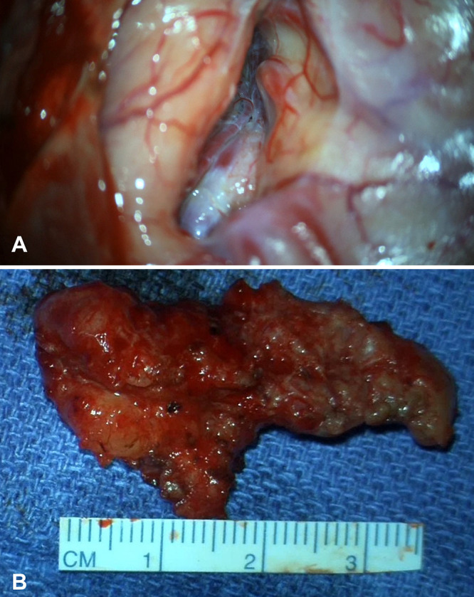
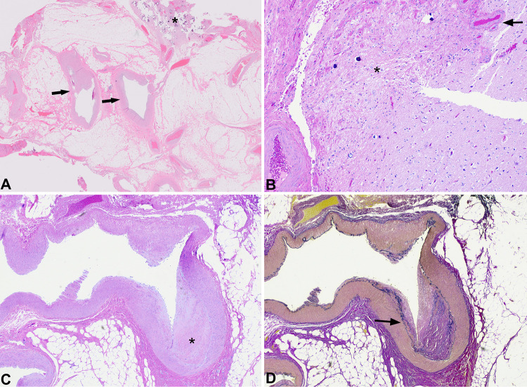
Observations: The authors present a case of heterotopic adipose tissue and a nonshunting arterial vascular malformation arising within a schizencephalic cleft in a 22-year-old male. Imaging showed right frontal gray matter abnormality and an associated suspected arteriovenous malformation with evidence of hemorrhage. Brain magnetic resonance imaging revealed right frontal polymicrogyria lining an open-lip schizencephaly, periventricular heterotopic gray matter, fat within the schizencephalic cleft, and gradient echo hypointensity concerning for prior hemorrhage. Histological assessment demonstrated mature adipose tissue with large-bore, thick-walled, irregular arteries. Mural calcifications and subendothelial cushions suggesting nonlaminar blood flow were observed. There were no arterialized veins or direct transitions from the arteries to veins. Hemosiderin deposition was scant, and hemorrhage was not present. The final diagnosis was consistent with ectopic mature adipose tissue and arteries with meningocerebral cicatrix.
Lessons: This example of a complex maldevelopment of derivatives of the meninx primitiva in association with cortical maldevelopment highlights the unique challenges from both a radiological and histological perspective during diagnostic workup.


 求助内容:
求助内容: 应助结果提醒方式:


