Muhammad Omer Altaf, Saad Khalil Chaudhry, Palwasha Gul, Waqas Ahmad, Atif Naveed, Islah Ud Din
{"title":"颅内轴外未分化多形性肉瘤;一份病例报告。","authors":"Muhammad Omer Altaf, Saad Khalil Chaudhry, Palwasha Gul, Waqas Ahmad, Atif Naveed, Islah Ud Din","doi":"10.37029/jcas.v6i2.357","DOIUrl":null,"url":null,"abstract":"<p><strong>Introduction: </strong>Head-and-neck sarcomas result in high mortality rates. A lot of new cases of sarcomas are diagnosed every year constituting about 1 % of all head-and-neck malignancies. Undifferentiated pleomorphic sarcomas (UPSs) are high-grade soft-tissue malignant tumours which occur primarily in limbs and retroperitoneal cavities. These tumours can often metastasize to the central nervous system. However, in rare instances, soft-tissue sarcomas may develop as a primary lesion within the intracranial compartments.</p><p><strong>Case description: </strong>A young male presented to the clinic with occipital headache and blurring of vision. Initial workup included brain contrast-enhanced computed tomography (CECT) and magnetic resonance imaging (MRI). The CECT suggested that there was an extra-axial mass present which was pressing against the adjacent left frontal lobe. Overlying frontal bone of the left side showed remodelling effect and associated mild periosteal reaction. MRI scan showed intracranial extra-axial lobulated mass with T1 intermediate to low-signal intensity and intermediate to high signals on T2 sequences. Heterogeneous enhancement on post-contrast sequences was also seen. The lesion had a broad-based attachment with dura mater and was closely applied to the orbital roof without orbital invasion. Staging positron emission tomography-CT scan showed a solitary site of disease in an intracranial location. Final diagnosis was confirmed by histopathology following excision of mass as UPS. Post-surgery MRI brain showed satisfactory post-operative appearance without any residual disease. The patient remained asymptomatic for 2 years and 6 months following the resection of the tumour.</p><p><strong>Practical implications: </strong>Most of the extra-axial intracranial soft-tissue tumours arise from the meninges with meningiomas making the substantial bulk; however, possibility of other relatively rare tumours of meningeal origin must not be ignored. Intracranial soft-tissue sarcomas mostly arise from meninges thus require a good understanding of clinical presentation as well as acquaintance with morphological features on radiological imaging to differentiate from other tumours. These can be treated with excision and radiotherapy along with sequential follow-ups to look for recurrence. Tissue sampling is mandatory followed by complete staging scan in case of sarcomas to rule out possible primary or secondary disease.</p>","PeriodicalId":73631,"journal":{"name":"Journal of cancer & allied specialties","volume":"6 2","pages":"e357"},"PeriodicalIF":0.0000,"publicationDate":"2020-05-12","publicationTypes":"Journal Article","fieldsOfStudy":null,"isOpenAccess":false,"openAccessPdf":"https://ftp.ncbi.nlm.nih.gov/pub/pmc/oa_pdf/cd/3e/JCAS-6-357.PMC10166348.pdf","citationCount":"0","resultStr":"{\"title\":\"Intracranial Extra-axial Undifferentiated Pleomorphic Sarcoma; a Case Report.\",\"authors\":\"Muhammad Omer Altaf, Saad Khalil Chaudhry, Palwasha Gul, Waqas Ahmad, Atif Naveed, Islah Ud Din\",\"doi\":\"10.37029/jcas.v6i2.357\",\"DOIUrl\":null,\"url\":null,\"abstract\":\"<p><strong>Introduction: </strong>Head-and-neck sarcomas result in high mortality rates. A lot of new cases of sarcomas are diagnosed every year constituting about 1 % of all head-and-neck malignancies. Undifferentiated pleomorphic sarcomas (UPSs) are high-grade soft-tissue malignant tumours which occur primarily in limbs and retroperitoneal cavities. These tumours can often metastasize to the central nervous system. However, in rare instances, soft-tissue sarcomas may develop as a primary lesion within the intracranial compartments.</p><p><strong>Case description: </strong>A young male presented to the clinic with occipital headache and blurring of vision. Initial workup included brain contrast-enhanced computed tomography (CECT) and magnetic resonance imaging (MRI). The CECT suggested that there was an extra-axial mass present which was pressing against the adjacent left frontal lobe. Overlying frontal bone of the left side showed remodelling effect and associated mild periosteal reaction. MRI scan showed intracranial extra-axial lobulated mass with T1 intermediate to low-signal intensity and intermediate to high signals on T2 sequences. Heterogeneous enhancement on post-contrast sequences was also seen. The lesion had a broad-based attachment with dura mater and was closely applied to the orbital roof without orbital invasion. Staging positron emission tomography-CT scan showed a solitary site of disease in an intracranial location. Final diagnosis was confirmed by histopathology following excision of mass as UPS. Post-surgery MRI brain showed satisfactory post-operative appearance without any residual disease. The patient remained asymptomatic for 2 years and 6 months following the resection of the tumour.</p><p><strong>Practical implications: </strong>Most of the extra-axial intracranial soft-tissue tumours arise from the meninges with meningiomas making the substantial bulk; however, possibility of other relatively rare tumours of meningeal origin must not be ignored. Intracranial soft-tissue sarcomas mostly arise from meninges thus require a good understanding of clinical presentation as well as acquaintance with morphological features on radiological imaging to differentiate from other tumours. These can be treated with excision and radiotherapy along with sequential follow-ups to look for recurrence. Tissue sampling is mandatory followed by complete staging scan in case of sarcomas to rule out possible primary or secondary disease.</p>\",\"PeriodicalId\":73631,\"journal\":{\"name\":\"Journal of cancer & allied specialties\",\"volume\":\"6 2\",\"pages\":\"e357\"},\"PeriodicalIF\":0.0000,\"publicationDate\":\"2020-05-12\",\"publicationTypes\":\"Journal Article\",\"fieldsOfStudy\":null,\"isOpenAccess\":false,\"openAccessPdf\":\"https://ftp.ncbi.nlm.nih.gov/pub/pmc/oa_pdf/cd/3e/JCAS-6-357.PMC10166348.pdf\",\"citationCount\":\"0\",\"resultStr\":null,\"platform\":\"Semanticscholar\",\"paperid\":null,\"PeriodicalName\":\"Journal of cancer & allied specialties\",\"FirstCategoryId\":\"1085\",\"ListUrlMain\":\"https://doi.org/10.37029/jcas.v6i2.357\",\"RegionNum\":0,\"RegionCategory\":null,\"ArticlePicture\":[],\"TitleCN\":null,\"AbstractTextCN\":null,\"PMCID\":null,\"EPubDate\":\"2020/1/1 0:00:00\",\"PubModel\":\"eCollection\",\"JCR\":\"\",\"JCRName\":\"\",\"Score\":null,\"Total\":0}","platform":"Semanticscholar","paperid":null,"PeriodicalName":"Journal of cancer & allied specialties","FirstCategoryId":"1085","ListUrlMain":"https://doi.org/10.37029/jcas.v6i2.357","RegionNum":0,"RegionCategory":null,"ArticlePicture":[],"TitleCN":null,"AbstractTextCN":null,"PMCID":null,"EPubDate":"2020/1/1 0:00:00","PubModel":"eCollection","JCR":"","JCRName":"","Score":null,"Total":0}
Intracranial Extra-axial Undifferentiated Pleomorphic Sarcoma; a Case Report.
Introduction: Head-and-neck sarcomas result in high mortality rates. A lot of new cases of sarcomas are diagnosed every year constituting about 1 % of all head-and-neck malignancies. Undifferentiated pleomorphic sarcomas (UPSs) are high-grade soft-tissue malignant tumours which occur primarily in limbs and retroperitoneal cavities. These tumours can often metastasize to the central nervous system. However, in rare instances, soft-tissue sarcomas may develop as a primary lesion within the intracranial compartments.
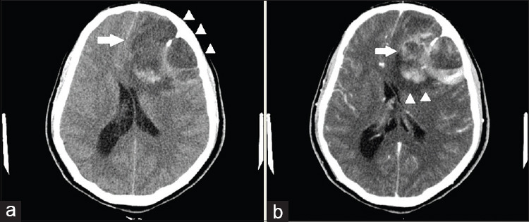
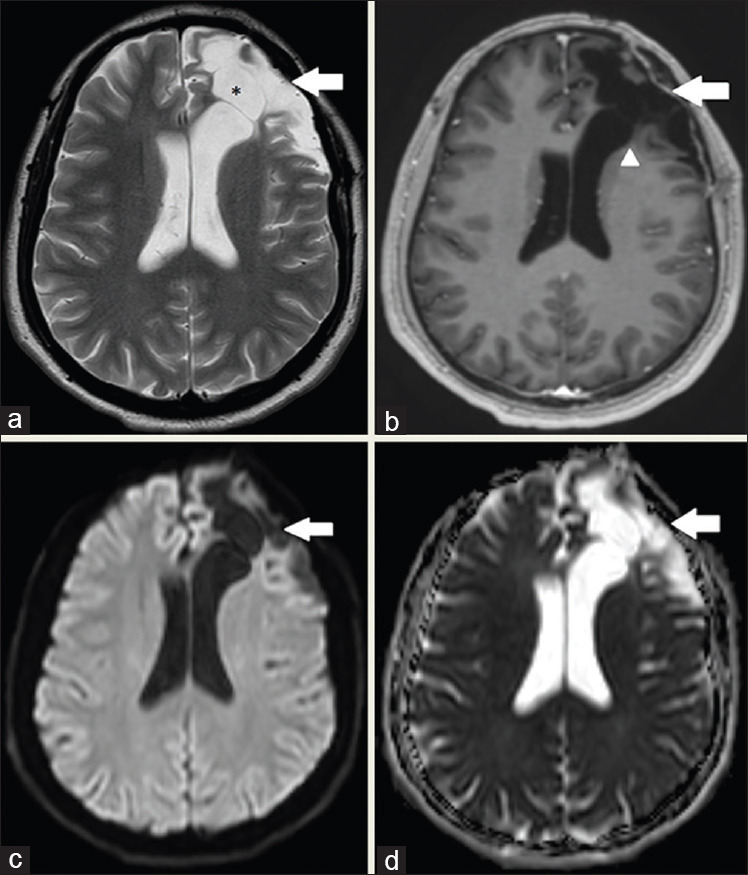
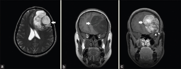
Case description: A young male presented to the clinic with occipital headache and blurring of vision. Initial workup included brain contrast-enhanced computed tomography (CECT) and magnetic resonance imaging (MRI). The CECT suggested that there was an extra-axial mass present which was pressing against the adjacent left frontal lobe. Overlying frontal bone of the left side showed remodelling effect and associated mild periosteal reaction. MRI scan showed intracranial extra-axial lobulated mass with T1 intermediate to low-signal intensity and intermediate to high signals on T2 sequences. Heterogeneous enhancement on post-contrast sequences was also seen. The lesion had a broad-based attachment with dura mater and was closely applied to the orbital roof without orbital invasion. Staging positron emission tomography-CT scan showed a solitary site of disease in an intracranial location. Final diagnosis was confirmed by histopathology following excision of mass as UPS. Post-surgery MRI brain showed satisfactory post-operative appearance without any residual disease. The patient remained asymptomatic for 2 years and 6 months following the resection of the tumour.
Practical implications: Most of the extra-axial intracranial soft-tissue tumours arise from the meninges with meningiomas making the substantial bulk; however, possibility of other relatively rare tumours of meningeal origin must not be ignored. Intracranial soft-tissue sarcomas mostly arise from meninges thus require a good understanding of clinical presentation as well as acquaintance with morphological features on radiological imaging to differentiate from other tumours. These can be treated with excision and radiotherapy along with sequential follow-ups to look for recurrence. Tissue sampling is mandatory followed by complete staging scan in case of sarcomas to rule out possible primary or secondary disease.

 求助内容:
求助内容: 应助结果提醒方式:


