Samir A A El-Gendy, Basma M Kamal, Mohamed A M Alsafy
{"title":"单峰骆驼(Camelus dromedarius)后趾骨和动脉的三维渲染体积CT重建。","authors":"Samir A A El-Gendy, Basma M Kamal, Mohamed A M Alsafy","doi":"10.1186/s40850-022-00151-8","DOIUrl":null,"url":null,"abstract":"<p><strong>Background: </strong>The 3D computed tomography produces detailed images of the digit bones in addition to the angiograph render volume 3D of the CT shows the relation between the arteries, bones, and tissues of the digit. Therefore, the present study used those imaging techniques to provide a complete description of the digit bones and arteries' origin, distribution, and course and their relations with surrounding structures in the Dromedary Camel. Which would serve as a guide for surgeons and students in distinguishing normal digit structures. The study used eight hind limbs of four adult camels of both sexes (two males and two females), aged 9-15 years (Mean ± SD, 11.80 ± 2.59 years). The samples were injected with latex with lead oxide were undergone 3D render volume CT (128-slice multi-detector CT scanning protocol) and angiography x-rays.</p><p><strong>Results: </strong>The blood vessels and correlated structures such as bones, tendons, and ligaments were examined using 3D CT in all directions, which was easier to view than angiography and dissected specimens. The arterial supply to the camel's hind foot was the A. digitalis plantaris communis III. The angiography render volume 3D of CT explained the blood supply of the bones and joints of digital regions and showed a good visualization of the many digit arteries. The metatarsals, the phalanges, and the sesamoid bones were visualized. A. plantaris medialis superficialis, A. digitalis plantaris communis III, A. digitalis plantaris communis II and IV, A. interdigitalis, rami articularis medialis and lateralis to the fetlock joint, ramus medialis and ramus lateralis of the A. digitalis plantaris communis III, A. digitalis plantaris propriae III et IV abaxialis, A. digitalis plantaris propriae III et IV axialis, Ramus phalangis axialis and abaxialis of the first phalanx, Ramus phalangis axialis and abaxialis of the second and third phalanges, and A. metatarsae plantaris III were visualized. The study discovered new blood vessel sources in dromedary camels, such as the ramus articularis to the fetlock and the ramus plantaris phalangis abaxialis proximalis and distalis of the first phalanx.</p><p><strong>Conclusions: </strong>The digital circulation angiography investigates the circulatory pattern of the camel hind digit, which can assist clinicians in diagnosing digit region affections. 3D CT explained improved visualization of bones and arteries, including many small branches in relation to surrounding structures, in some views better than others.</p>","PeriodicalId":48590,"journal":{"name":"BMC Zoology","volume":"7 1","pages":"49"},"PeriodicalIF":1.7000,"publicationDate":"2022-08-31","publicationTypes":"Journal Article","fieldsOfStudy":null,"isOpenAccess":false,"openAccessPdf":"https://www.ncbi.nlm.nih.gov/pmc/articles/PMC10127028/pdf/","citationCount":"2","resultStr":"{\"title\":\"3D render volume CT reconstruction of the bones and arteries of the hind digit of the dromedary camel (Camelus dromedarius).\",\"authors\":\"Samir A A El-Gendy, Basma M Kamal, Mohamed A M Alsafy\",\"doi\":\"10.1186/s40850-022-00151-8\",\"DOIUrl\":null,\"url\":null,\"abstract\":\"<p><strong>Background: </strong>The 3D computed tomography produces detailed images of the digit bones in addition to the angiograph render volume 3D of the CT shows the relation between the arteries, bones, and tissues of the digit. Therefore, the present study used those imaging techniques to provide a complete description of the digit bones and arteries' origin, distribution, and course and their relations with surrounding structures in the Dromedary Camel. Which would serve as a guide for surgeons and students in distinguishing normal digit structures. The study used eight hind limbs of four adult camels of both sexes (two males and two females), aged 9-15 years (Mean ± SD, 11.80 ± 2.59 years). The samples were injected with latex with lead oxide were undergone 3D render volume CT (128-slice multi-detector CT scanning protocol) and angiography x-rays.</p><p><strong>Results: </strong>The blood vessels and correlated structures such as bones, tendons, and ligaments were examined using 3D CT in all directions, which was easier to view than angiography and dissected specimens. The arterial supply to the camel's hind foot was the A. digitalis plantaris communis III. The angiography render volume 3D of CT explained the blood supply of the bones and joints of digital regions and showed a good visualization of the many digit arteries. The metatarsals, the phalanges, and the sesamoid bones were visualized. A. plantaris medialis superficialis, A. digitalis plantaris communis III, A. digitalis plantaris communis II and IV, A. interdigitalis, rami articularis medialis and lateralis to the fetlock joint, ramus medialis and ramus lateralis of the A. digitalis plantaris communis III, A. digitalis plantaris propriae III et IV abaxialis, A. digitalis plantaris propriae III et IV axialis, Ramus phalangis axialis and abaxialis of the first phalanx, Ramus phalangis axialis and abaxialis of the second and third phalanges, and A. metatarsae plantaris III were visualized. The study discovered new blood vessel sources in dromedary camels, such as the ramus articularis to the fetlock and the ramus plantaris phalangis abaxialis proximalis and distalis of the first phalanx.</p><p><strong>Conclusions: </strong>The digital circulation angiography investigates the circulatory pattern of the camel hind digit, which can assist clinicians in diagnosing digit region affections. 3D CT explained improved visualization of bones and arteries, including many small branches in relation to surrounding structures, in some views better than others.</p>\",\"PeriodicalId\":48590,\"journal\":{\"name\":\"BMC Zoology\",\"volume\":\"7 1\",\"pages\":\"49\"},\"PeriodicalIF\":1.7000,\"publicationDate\":\"2022-08-31\",\"publicationTypes\":\"Journal Article\",\"fieldsOfStudy\":null,\"isOpenAccess\":false,\"openAccessPdf\":\"https://www.ncbi.nlm.nih.gov/pmc/articles/PMC10127028/pdf/\",\"citationCount\":\"2\",\"resultStr\":null,\"platform\":\"Semanticscholar\",\"paperid\":null,\"PeriodicalName\":\"BMC Zoology\",\"FirstCategoryId\":\"99\",\"ListUrlMain\":\"https://doi.org/10.1186/s40850-022-00151-8\",\"RegionNum\":3,\"RegionCategory\":\"生物学\",\"ArticlePicture\":[],\"TitleCN\":null,\"AbstractTextCN\":null,\"PMCID\":null,\"EPubDate\":\"\",\"PubModel\":\"\",\"JCR\":\"Q2\",\"JCRName\":\"ZOOLOGY\",\"Score\":null,\"Total\":0}","platform":"Semanticscholar","paperid":null,"PeriodicalName":"BMC Zoology","FirstCategoryId":"99","ListUrlMain":"https://doi.org/10.1186/s40850-022-00151-8","RegionNum":3,"RegionCategory":"生物学","ArticlePicture":[],"TitleCN":null,"AbstractTextCN":null,"PMCID":null,"EPubDate":"","PubModel":"","JCR":"Q2","JCRName":"ZOOLOGY","Score":null,"Total":0}
3D render volume CT reconstruction of the bones and arteries of the hind digit of the dromedary camel (Camelus dromedarius).
Background: The 3D computed tomography produces detailed images of the digit bones in addition to the angiograph render volume 3D of the CT shows the relation between the arteries, bones, and tissues of the digit. Therefore, the present study used those imaging techniques to provide a complete description of the digit bones and arteries' origin, distribution, and course and their relations with surrounding structures in the Dromedary Camel. Which would serve as a guide for surgeons and students in distinguishing normal digit structures. The study used eight hind limbs of four adult camels of both sexes (two males and two females), aged 9-15 years (Mean ± SD, 11.80 ± 2.59 years). The samples were injected with latex with lead oxide were undergone 3D render volume CT (128-slice multi-detector CT scanning protocol) and angiography x-rays.
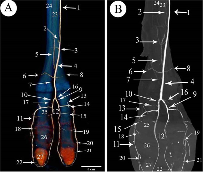
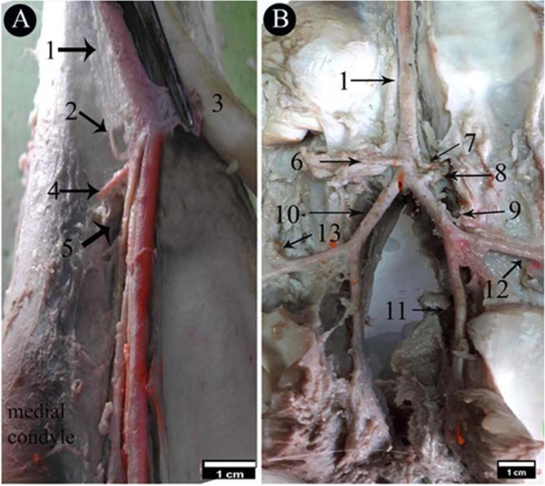
Results: The blood vessels and correlated structures such as bones, tendons, and ligaments were examined using 3D CT in all directions, which was easier to view than angiography and dissected specimens. The arterial supply to the camel's hind foot was the A. digitalis plantaris communis III. The angiography render volume 3D of CT explained the blood supply of the bones and joints of digital regions and showed a good visualization of the many digit arteries. The metatarsals, the phalanges, and the sesamoid bones were visualized. A. plantaris medialis superficialis, A. digitalis plantaris communis III, A. digitalis plantaris communis II and IV, A. interdigitalis, rami articularis medialis and lateralis to the fetlock joint, ramus medialis and ramus lateralis of the A. digitalis plantaris communis III, A. digitalis plantaris propriae III et IV abaxialis, A. digitalis plantaris propriae III et IV axialis, Ramus phalangis axialis and abaxialis of the first phalanx, Ramus phalangis axialis and abaxialis of the second and third phalanges, and A. metatarsae plantaris III were visualized. The study discovered new blood vessel sources in dromedary camels, such as the ramus articularis to the fetlock and the ramus plantaris phalangis abaxialis proximalis and distalis of the first phalanx.
Conclusions: The digital circulation angiography investigates the circulatory pattern of the camel hind digit, which can assist clinicians in diagnosing digit region affections. 3D CT explained improved visualization of bones and arteries, including many small branches in relation to surrounding structures, in some views better than others.
BMC ZoologyAgricultural and Biological Sciences-Animal Science and Zoology
CiteScore
2.30
自引率
6.20%
发文量
53
审稿时长
24 weeks
期刊介绍:
BMC Zoology is an open access, peer-reviewed journal that considers articles on all aspects of zoology, including physiology, mechanistic and functional studies, anatomy, life history, behavior, signalling and communication, cognition, parasitism, taxonomy and conservation.

 求助内容:
求助内容: 应助结果提醒方式:


