Albina Aldomà-Balasch, Ma Isabel Hernández-Martín, Ma Dolors Viles-Bertran
{"title":"年轻运动员的t波倒置:正常还是病态?","authors":"Albina Aldomà-Balasch, Ma Isabel Hernández-Martín, Ma Dolors Viles-Bertran","doi":"10.24875/ACM.210003261","DOIUrl":null,"url":null,"abstract":"A 17-years-old white male patient with no personal history of interest and asymptomatic from the cardiovascular point of view, but with a family history of paternal grandfather with dilated cardiomyopathy and mild depressed left ventricle ejection fraction (LVEF) who died at 83 years of age of a noncardiological cause. In an electrocardiogram (ECG) performed during a sports examination (field hockey player), T-wave inversion (TWI) was identified in the inferior leads (Fig. 1). For further evaluation, an echocardiogram was performed, showing a pattern of hypertrabeculation in the inferior, lateral, and apical walls of the left ventricular, which was confirmed by cardiac magnetic resonance imaging (Fig. 2A and 2B), fulfilling the criteria for noncompaction, with no other notable findings. As a result of these findings, the 57-year-old boy’s father, who was asymptomatic cardio-vascular and had a non-pathological ECG, was also evaluated and showed noncompaction cardiomyopathy with normal LVEF.","PeriodicalId":8360,"journal":{"name":"Archivos de cardiologia de Mexico","volume":"93 1","pages":"096-097"},"PeriodicalIF":0.7000,"publicationDate":"2023-02-02","publicationTypes":"Journal Article","fieldsOfStudy":null,"isOpenAccess":false,"openAccessPdf":"https://ftp.ncbi.nlm.nih.gov/pub/pmc/oa_pdf/8d/05/7567AX221-ACM-93-96.PMC10161837.pdf","citationCount":"0","resultStr":"{\"title\":\"T-wave inversion in young athletes: Normal or pathological?\",\"authors\":\"Albina Aldomà-Balasch, Ma Isabel Hernández-Martín, Ma Dolors Viles-Bertran\",\"doi\":\"10.24875/ACM.210003261\",\"DOIUrl\":null,\"url\":null,\"abstract\":\"A 17-years-old white male patient with no personal history of interest and asymptomatic from the cardiovascular point of view, but with a family history of paternal grandfather with dilated cardiomyopathy and mild depressed left ventricle ejection fraction (LVEF) who died at 83 years of age of a noncardiological cause. In an electrocardiogram (ECG) performed during a sports examination (field hockey player), T-wave inversion (TWI) was identified in the inferior leads (Fig. 1). For further evaluation, an echocardiogram was performed, showing a pattern of hypertrabeculation in the inferior, lateral, and apical walls of the left ventricular, which was confirmed by cardiac magnetic resonance imaging (Fig. 2A and 2B), fulfilling the criteria for noncompaction, with no other notable findings. As a result of these findings, the 57-year-old boy’s father, who was asymptomatic cardio-vascular and had a non-pathological ECG, was also evaluated and showed noncompaction cardiomyopathy with normal LVEF.\",\"PeriodicalId\":8360,\"journal\":{\"name\":\"Archivos de cardiologia de Mexico\",\"volume\":\"93 1\",\"pages\":\"096-097\"},\"PeriodicalIF\":0.7000,\"publicationDate\":\"2023-02-02\",\"publicationTypes\":\"Journal Article\",\"fieldsOfStudy\":null,\"isOpenAccess\":false,\"openAccessPdf\":\"https://ftp.ncbi.nlm.nih.gov/pub/pmc/oa_pdf/8d/05/7567AX221-ACM-93-96.PMC10161837.pdf\",\"citationCount\":\"0\",\"resultStr\":null,\"platform\":\"Semanticscholar\",\"paperid\":null,\"PeriodicalName\":\"Archivos de cardiologia de Mexico\",\"FirstCategoryId\":\"1085\",\"ListUrlMain\":\"https://doi.org/10.24875/ACM.210003261\",\"RegionNum\":0,\"RegionCategory\":null,\"ArticlePicture\":[],\"TitleCN\":null,\"AbstractTextCN\":null,\"PMCID\":null,\"EPubDate\":\"\",\"PubModel\":\"\",\"JCR\":\"Q4\",\"JCRName\":\"CARDIAC & CARDIOVASCULAR SYSTEMS\",\"Score\":null,\"Total\":0}","platform":"Semanticscholar","paperid":null,"PeriodicalName":"Archivos de cardiologia de Mexico","FirstCategoryId":"1085","ListUrlMain":"https://doi.org/10.24875/ACM.210003261","RegionNum":0,"RegionCategory":null,"ArticlePicture":[],"TitleCN":null,"AbstractTextCN":null,"PMCID":null,"EPubDate":"","PubModel":"","JCR":"Q4","JCRName":"CARDIAC & CARDIOVASCULAR SYSTEMS","Score":null,"Total":0}
T-wave inversion in young athletes: Normal or pathological?
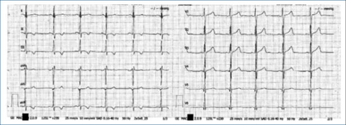
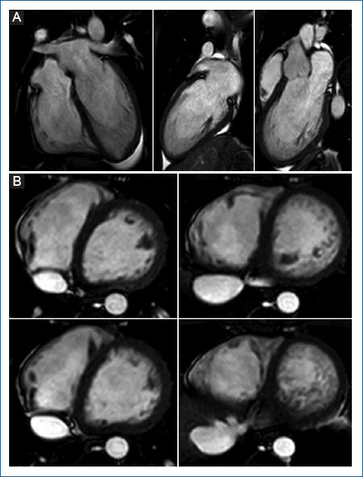
A 17-years-old white male patient with no personal history of interest and asymptomatic from the cardiovascular point of view, but with a family history of paternal grandfather with dilated cardiomyopathy and mild depressed left ventricle ejection fraction (LVEF) who died at 83 years of age of a noncardiological cause. In an electrocardiogram (ECG) performed during a sports examination (field hockey player), T-wave inversion (TWI) was identified in the inferior leads (Fig. 1). For further evaluation, an echocardiogram was performed, showing a pattern of hypertrabeculation in the inferior, lateral, and apical walls of the left ventricular, which was confirmed by cardiac magnetic resonance imaging (Fig. 2A and 2B), fulfilling the criteria for noncompaction, with no other notable findings. As a result of these findings, the 57-year-old boy’s father, who was asymptomatic cardio-vascular and had a non-pathological ECG, was also evaluated and showed noncompaction cardiomyopathy with normal LVEF.

 求助内容:
求助内容: 应助结果提醒方式:


