Mohamed Nasr, Suhaib Alsayed Naeem, Ibrahim El-Shenbaby, Fatma Mahmoud Abdelraheem Mohamed, Safinaz Moustafa Mahmoud, Tamer M M Abuamara, Wagih M Abd-Elhay, Fayez Mohammed Abd Elfattah Elbayoumy, Ahmad Elkot, Tarek Shikhon, Mostafa Abo-Akrab, Mohamed A Doma, Abdulkarim Hasan
{"title":"石榴籽和石榴皮乙醇提取物的抗癌潜力及其相关的遗传、组织学、免疫组织化学、凋亡和氧化应激谱:体外研究。","authors":"Mohamed Nasr, Suhaib Alsayed Naeem, Ibrahim El-Shenbaby, Fatma Mahmoud Abdelraheem Mohamed, Safinaz Moustafa Mahmoud, Tamer M M Abuamara, Wagih M Abd-Elhay, Fayez Mohammed Abd Elfattah Elbayoumy, Ahmad Elkot, Tarek Shikhon, Mostafa Abo-Akrab, Mohamed A Doma, Abdulkarim Hasan","doi":"10.2147/JEP.S404321","DOIUrl":null,"url":null,"abstract":"<p><strong>Introduction: </strong>Owing to their great quantity of hydrolyzable anthocyanins and tannins, the peel and seeds of pomegranate are edible and possess potent anti-oxidant and anti-inflammatory characteristics. This work aims to trace the pomegranate seed and peel ethanolic extracts' anticancer activity against liver cancer cell line, namely HepG2 and related histopathological, immunohistochemical, genetic and oxidative stress profile.</p><p><strong>Methods: </strong>In vitro study for both seed and peel extract showed the prevalence of phenols, polyphenols and acids, those have anti-proliferative potential against liver cancer cell line (HepG2) with 50% inhibitory concentration (IC50) of seed significantly reduced that of peel. Toxicity of test extracts was concentration dependent and accompanied with cell cycle arrest and cell death at theG0/G1 and S phases but not at the G2/M phase. Cell arrest was supplemented with raised ROS, MDA and decreased SOD, GSH and Catalase.</p><p><strong>Results and discussion: </strong>Apoptosis-related genes showed significant up-expression of pro-apoptotic gene (<i>P53</i>), <i>Cy-C</i>, <i>Bax</i>, and <i>casp-3</i> and down expression of anti-apoptotic gene (<i>Bcl-2</i>). Also, Casp-3 and P53 proteins were substantially expressed under the effect of test extracts. Histopathological study demonstrated that the untreated cells (control group) were regular cells with nuclear pleomorphism and hyperchromatic nuclei, while seed and peel extracts-treated cells showed necrosis, mixed euchromatin and heterochromatin, intra-nuclear eosinophilic structures, burst cell membranes, and the shrunken apoptotic cells with nuclear membranes and irregular cells. Finally, <i>PCNA</i> gene detected by immunohistochemistry was down regulated significantly under the effect of seed extract treatment than in case of cell medication with peel extract.</p>","PeriodicalId":15846,"journal":{"name":"Journal of Experimental Pharmacology","volume":"15 ","pages":"191-205"},"PeriodicalIF":0.0000,"publicationDate":"2023-01-01","publicationTypes":"Journal Article","fieldsOfStudy":null,"isOpenAccess":false,"openAccessPdf":"https://ftp.ncbi.nlm.nih.gov/pub/pmc/oa_pdf/04/fb/jep-15-191.PMC10115208.pdf","citationCount":"4","resultStr":"{\"title\":\"Pomegranate Seeds and Peel Ethanolic Extracts Anticancer Potentials and Related Genetic, Histological, Immunohistochemical, Apoptotic and Oxidative Stress Profiles: In vitro Study.\",\"authors\":\"Mohamed Nasr, Suhaib Alsayed Naeem, Ibrahim El-Shenbaby, Fatma Mahmoud Abdelraheem Mohamed, Safinaz Moustafa Mahmoud, Tamer M M Abuamara, Wagih M Abd-Elhay, Fayez Mohammed Abd Elfattah Elbayoumy, Ahmad Elkot, Tarek Shikhon, Mostafa Abo-Akrab, Mohamed A Doma, Abdulkarim Hasan\",\"doi\":\"10.2147/JEP.S404321\",\"DOIUrl\":null,\"url\":null,\"abstract\":\"<p><strong>Introduction: </strong>Owing to their great quantity of hydrolyzable anthocyanins and tannins, the peel and seeds of pomegranate are edible and possess potent anti-oxidant and anti-inflammatory characteristics. This work aims to trace the pomegranate seed and peel ethanolic extracts' anticancer activity against liver cancer cell line, namely HepG2 and related histopathological, immunohistochemical, genetic and oxidative stress profile.</p><p><strong>Methods: </strong>In vitro study for both seed and peel extract showed the prevalence of phenols, polyphenols and acids, those have anti-proliferative potential against liver cancer cell line (HepG2) with 50% inhibitory concentration (IC50) of seed significantly reduced that of peel. Toxicity of test extracts was concentration dependent and accompanied with cell cycle arrest and cell death at theG0/G1 and S phases but not at the G2/M phase. Cell arrest was supplemented with raised ROS, MDA and decreased SOD, GSH and Catalase.</p><p><strong>Results and discussion: </strong>Apoptosis-related genes showed significant up-expression of pro-apoptotic gene (<i>P53</i>), <i>Cy-C</i>, <i>Bax</i>, and <i>casp-3</i> and down expression of anti-apoptotic gene (<i>Bcl-2</i>). Also, Casp-3 and P53 proteins were substantially expressed under the effect of test extracts. Histopathological study demonstrated that the untreated cells (control group) were regular cells with nuclear pleomorphism and hyperchromatic nuclei, while seed and peel extracts-treated cells showed necrosis, mixed euchromatin and heterochromatin, intra-nuclear eosinophilic structures, burst cell membranes, and the shrunken apoptotic cells with nuclear membranes and irregular cells. Finally, <i>PCNA</i> gene detected by immunohistochemistry was down regulated significantly under the effect of seed extract treatment than in case of cell medication with peel extract.</p>\",\"PeriodicalId\":15846,\"journal\":{\"name\":\"Journal of Experimental Pharmacology\",\"volume\":\"15 \",\"pages\":\"191-205\"},\"PeriodicalIF\":0.0000,\"publicationDate\":\"2023-01-01\",\"publicationTypes\":\"Journal Article\",\"fieldsOfStudy\":null,\"isOpenAccess\":false,\"openAccessPdf\":\"https://ftp.ncbi.nlm.nih.gov/pub/pmc/oa_pdf/04/fb/jep-15-191.PMC10115208.pdf\",\"citationCount\":\"4\",\"resultStr\":null,\"platform\":\"Semanticscholar\",\"paperid\":null,\"PeriodicalName\":\"Journal of Experimental Pharmacology\",\"FirstCategoryId\":\"1085\",\"ListUrlMain\":\"https://doi.org/10.2147/JEP.S404321\",\"RegionNum\":0,\"RegionCategory\":null,\"ArticlePicture\":[],\"TitleCN\":null,\"AbstractTextCN\":null,\"PMCID\":null,\"EPubDate\":\"\",\"PubModel\":\"\",\"JCR\":\"Q2\",\"JCRName\":\"Medicine\",\"Score\":null,\"Total\":0}","platform":"Semanticscholar","paperid":null,"PeriodicalName":"Journal of Experimental Pharmacology","FirstCategoryId":"1085","ListUrlMain":"https://doi.org/10.2147/JEP.S404321","RegionNum":0,"RegionCategory":null,"ArticlePicture":[],"TitleCN":null,"AbstractTextCN":null,"PMCID":null,"EPubDate":"","PubModel":"","JCR":"Q2","JCRName":"Medicine","Score":null,"Total":0}
Pomegranate Seeds and Peel Ethanolic Extracts Anticancer Potentials and Related Genetic, Histological, Immunohistochemical, Apoptotic and Oxidative Stress Profiles: In vitro Study.
Introduction: Owing to their great quantity of hydrolyzable anthocyanins and tannins, the peel and seeds of pomegranate are edible and possess potent anti-oxidant and anti-inflammatory characteristics. This work aims to trace the pomegranate seed and peel ethanolic extracts' anticancer activity against liver cancer cell line, namely HepG2 and related histopathological, immunohistochemical, genetic and oxidative stress profile.
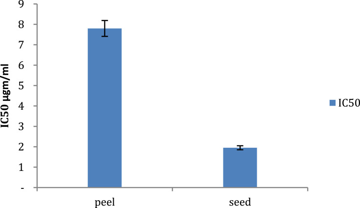
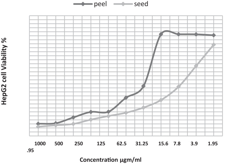
Methods: In vitro study for both seed and peel extract showed the prevalence of phenols, polyphenols and acids, those have anti-proliferative potential against liver cancer cell line (HepG2) with 50% inhibitory concentration (IC50) of seed significantly reduced that of peel. Toxicity of test extracts was concentration dependent and accompanied with cell cycle arrest and cell death at theG0/G1 and S phases but not at the G2/M phase. Cell arrest was supplemented with raised ROS, MDA and decreased SOD, GSH and Catalase.
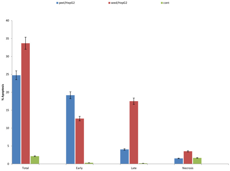
Results and discussion: Apoptosis-related genes showed significant up-expression of pro-apoptotic gene (P53), Cy-C, Bax, and casp-3 and down expression of anti-apoptotic gene (Bcl-2). Also, Casp-3 and P53 proteins were substantially expressed under the effect of test extracts. Histopathological study demonstrated that the untreated cells (control group) were regular cells with nuclear pleomorphism and hyperchromatic nuclei, while seed and peel extracts-treated cells showed necrosis, mixed euchromatin and heterochromatin, intra-nuclear eosinophilic structures, burst cell membranes, and the shrunken apoptotic cells with nuclear membranes and irregular cells. Finally, PCNA gene detected by immunohistochemistry was down regulated significantly under the effect of seed extract treatment than in case of cell medication with peel extract.

 求助内容:
求助内容: 应助结果提醒方式:


