Till D Lerch, Tilman Kaim, Markus S Hanke, Florian Schmaranzer, Simon D Steppacher, Jasmin D Busch, Eduardo N Novais, Kai Ziebarth
{"title":"股骨骨骺滑动患者术前髋关节磁共振成像对股骨后翻的评估:对髋关节撞击风险评估的理论意义。","authors":"Till D Lerch, Tilman Kaim, Markus S Hanke, Florian Schmaranzer, Simon D Steppacher, Jasmin D Busch, Eduardo N Novais, Kai Ziebarth","doi":"10.1177/18632521221149044","DOIUrl":null,"url":null,"abstract":"<p><strong>Purpose: </strong>Slipped capital femoral epiphysis is a common pediatric hip disease and was associated with femoral retroversion, but femoral version was rarely measured. Therefore, mean femoral version, mean femoral neck version, and prevalence of femoral retroversion were analyzed for slipped capital femoral epiphysis patients.</p><p><strong>Methods: </strong>A retrospective observational study evaluating preoperative hip magnetic resonance imaging of 27 patients (49 hips) was performed. Twenty-seven untreated slipped capital femoral epiphysis patients (28 slipped capital femoral epiphysis hips and 21 contralateral hips, age 10-16 years) were evaluated (79% stable slipped capital femoral epiphysis, 22 patients; 43% severe slipped capital femoral epiphysis, 12 patients). Femoral version was measured using Murphy method on magnetic resonance imaging (January 2014-December 2021, rapid bilateral 3-dimensional T1 water-only Dixon-based images of pelvis and knee). All slipped capital femoral epiphysis patients underwent surgery after magnetic resonance imaging.</p><p><strong>Results: </strong>Mean femoral version of slipped capital femoral epiphysis patients (-1° ± 15°) was significantly (p < 0.001) lower compared to contralateral side (15° ± 14°). Femoral version of slipped capital femoral epiphysis patients had significantly (p < 0.001) wider range from -42° to 35° (range 77°) compared to contralateral side (-5° to 44°, range 49°). Mean femoral neck version of slipped capital femoral epiphysis patients (6° ± 15°) was lower compared to contralateral side (11° ± 12°). Fifteen slipped capital femoral epiphysis patients (54%) had absolute femoral retroversion (femoral version < 0°). Six of the 12 hips (50%) with severe slips and 4 of the 8 hips (50%) with mild slips had absolute femoral retroversion (femoral version < 0°). Ten slipped capital femoral epiphysis patients (40%) had absolute femoral neck retroversion (femoral neck version < 0°).</p><p><strong>Conclusion: </strong>Although slipped capital femoral epiphysis patients showed asymmetrically lower femoral version compared to contralateral side, there was a wide range of femoral version, underlining the importance of patient-specific femoral version analysis on preoperative magnetic resonance imaging. Absolute femoral retroversion was prevalent in half of slipped capital femoral epiphysis patients, in half of severe slipped capital femoral epiphysis patients, and in half of mild slipped capital femoral epiphysis patients. This has implications for anterior hip impingement and for surgical treatment with in situ pinning or femoral osteotomy (e.g. proximal femoral derotation osteotomy) or other hip preservation surgery.</p>","PeriodicalId":56060,"journal":{"name":"Journal of Childrens Orthopaedics","volume":"17 2","pages":"116-125"},"PeriodicalIF":1.3000,"publicationDate":"2023-04-01","publicationTypes":"Journal Article","fieldsOfStudy":null,"isOpenAccess":false,"openAccessPdf":"https://ftp.ncbi.nlm.nih.gov/pub/pmc/oa_pdf/ea/98/10.1177_18632521221149044.PMC10080244.pdf","citationCount":"1","resultStr":"{\"title\":\"Assessment of femoral retroversion on preoperative hip magnetic resonance imaging in patients with slipped capital femoral epiphysis: Theoretical implications for hip impingement risk estimation.\",\"authors\":\"Till D Lerch, Tilman Kaim, Markus S Hanke, Florian Schmaranzer, Simon D Steppacher, Jasmin D Busch, Eduardo N Novais, Kai Ziebarth\",\"doi\":\"10.1177/18632521221149044\",\"DOIUrl\":null,\"url\":null,\"abstract\":\"<p><strong>Purpose: </strong>Slipped capital femoral epiphysis is a common pediatric hip disease and was associated with femoral retroversion, but femoral version was rarely measured. Therefore, mean femoral version, mean femoral neck version, and prevalence of femoral retroversion were analyzed for slipped capital femoral epiphysis patients.</p><p><strong>Methods: </strong>A retrospective observational study evaluating preoperative hip magnetic resonance imaging of 27 patients (49 hips) was performed. Twenty-seven untreated slipped capital femoral epiphysis patients (28 slipped capital femoral epiphysis hips and 21 contralateral hips, age 10-16 years) were evaluated (79% stable slipped capital femoral epiphysis, 22 patients; 43% severe slipped capital femoral epiphysis, 12 patients). Femoral version was measured using Murphy method on magnetic resonance imaging (January 2014-December 2021, rapid bilateral 3-dimensional T1 water-only Dixon-based images of pelvis and knee). All slipped capital femoral epiphysis patients underwent surgery after magnetic resonance imaging.</p><p><strong>Results: </strong>Mean femoral version of slipped capital femoral epiphysis patients (-1° ± 15°) was significantly (p < 0.001) lower compared to contralateral side (15° ± 14°). Femoral version of slipped capital femoral epiphysis patients had significantly (p < 0.001) wider range from -42° to 35° (range 77°) compared to contralateral side (-5° to 44°, range 49°). Mean femoral neck version of slipped capital femoral epiphysis patients (6° ± 15°) was lower compared to contralateral side (11° ± 12°). Fifteen slipped capital femoral epiphysis patients (54%) had absolute femoral retroversion (femoral version < 0°). Six of the 12 hips (50%) with severe slips and 4 of the 8 hips (50%) with mild slips had absolute femoral retroversion (femoral version < 0°). Ten slipped capital femoral epiphysis patients (40%) had absolute femoral neck retroversion (femoral neck version < 0°).</p><p><strong>Conclusion: </strong>Although slipped capital femoral epiphysis patients showed asymmetrically lower femoral version compared to contralateral side, there was a wide range of femoral version, underlining the importance of patient-specific femoral version analysis on preoperative magnetic resonance imaging. Absolute femoral retroversion was prevalent in half of slipped capital femoral epiphysis patients, in half of severe slipped capital femoral epiphysis patients, and in half of mild slipped capital femoral epiphysis patients. This has implications for anterior hip impingement and for surgical treatment with in situ pinning or femoral osteotomy (e.g. proximal femoral derotation osteotomy) or other hip preservation surgery.</p>\",\"PeriodicalId\":56060,\"journal\":{\"name\":\"Journal of Childrens Orthopaedics\",\"volume\":\"17 2\",\"pages\":\"116-125\"},\"PeriodicalIF\":1.3000,\"publicationDate\":\"2023-04-01\",\"publicationTypes\":\"Journal Article\",\"fieldsOfStudy\":null,\"isOpenAccess\":false,\"openAccessPdf\":\"https://ftp.ncbi.nlm.nih.gov/pub/pmc/oa_pdf/ea/98/10.1177_18632521221149044.PMC10080244.pdf\",\"citationCount\":\"1\",\"resultStr\":null,\"platform\":\"Semanticscholar\",\"paperid\":null,\"PeriodicalName\":\"Journal of Childrens Orthopaedics\",\"FirstCategoryId\":\"3\",\"ListUrlMain\":\"https://doi.org/10.1177/18632521221149044\",\"RegionNum\":4,\"RegionCategory\":\"医学\",\"ArticlePicture\":[],\"TitleCN\":null,\"AbstractTextCN\":null,\"PMCID\":null,\"EPubDate\":\"\",\"PubModel\":\"\",\"JCR\":\"Q3\",\"JCRName\":\"ORTHOPEDICS\",\"Score\":null,\"Total\":0}","platform":"Semanticscholar","paperid":null,"PeriodicalName":"Journal of Childrens Orthopaedics","FirstCategoryId":"3","ListUrlMain":"https://doi.org/10.1177/18632521221149044","RegionNum":4,"RegionCategory":"医学","ArticlePicture":[],"TitleCN":null,"AbstractTextCN":null,"PMCID":null,"EPubDate":"","PubModel":"","JCR":"Q3","JCRName":"ORTHOPEDICS","Score":null,"Total":0}
Assessment of femoral retroversion on preoperative hip magnetic resonance imaging in patients with slipped capital femoral epiphysis: Theoretical implications for hip impingement risk estimation.
Purpose: Slipped capital femoral epiphysis is a common pediatric hip disease and was associated with femoral retroversion, but femoral version was rarely measured. Therefore, mean femoral version, mean femoral neck version, and prevalence of femoral retroversion were analyzed for slipped capital femoral epiphysis patients.
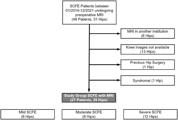
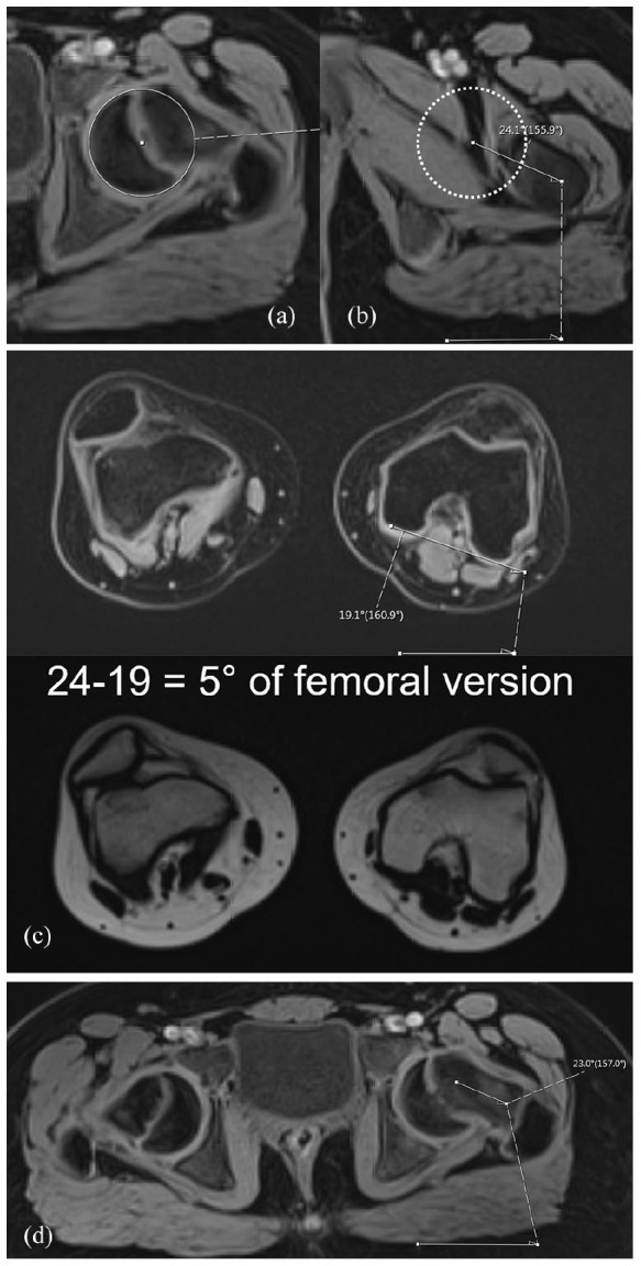
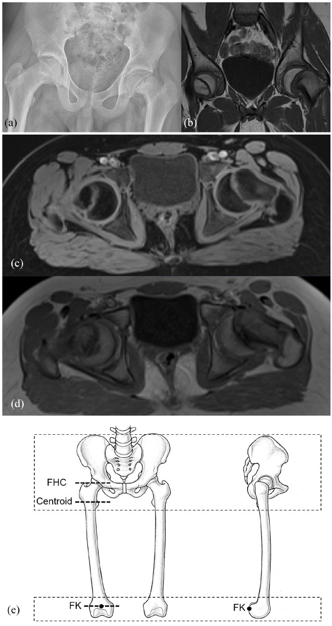
Methods: A retrospective observational study evaluating preoperative hip magnetic resonance imaging of 27 patients (49 hips) was performed. Twenty-seven untreated slipped capital femoral epiphysis patients (28 slipped capital femoral epiphysis hips and 21 contralateral hips, age 10-16 years) were evaluated (79% stable slipped capital femoral epiphysis, 22 patients; 43% severe slipped capital femoral epiphysis, 12 patients). Femoral version was measured using Murphy method on magnetic resonance imaging (January 2014-December 2021, rapid bilateral 3-dimensional T1 water-only Dixon-based images of pelvis and knee). All slipped capital femoral epiphysis patients underwent surgery after magnetic resonance imaging.
Results: Mean femoral version of slipped capital femoral epiphysis patients (-1° ± 15°) was significantly (p < 0.001) lower compared to contralateral side (15° ± 14°). Femoral version of slipped capital femoral epiphysis patients had significantly (p < 0.001) wider range from -42° to 35° (range 77°) compared to contralateral side (-5° to 44°, range 49°). Mean femoral neck version of slipped capital femoral epiphysis patients (6° ± 15°) was lower compared to contralateral side (11° ± 12°). Fifteen slipped capital femoral epiphysis patients (54%) had absolute femoral retroversion (femoral version < 0°). Six of the 12 hips (50%) with severe slips and 4 of the 8 hips (50%) with mild slips had absolute femoral retroversion (femoral version < 0°). Ten slipped capital femoral epiphysis patients (40%) had absolute femoral neck retroversion (femoral neck version < 0°).
Conclusion: Although slipped capital femoral epiphysis patients showed asymmetrically lower femoral version compared to contralateral side, there was a wide range of femoral version, underlining the importance of patient-specific femoral version analysis on preoperative magnetic resonance imaging. Absolute femoral retroversion was prevalent in half of slipped capital femoral epiphysis patients, in half of severe slipped capital femoral epiphysis patients, and in half of mild slipped capital femoral epiphysis patients. This has implications for anterior hip impingement and for surgical treatment with in situ pinning or femoral osteotomy (e.g. proximal femoral derotation osteotomy) or other hip preservation surgery.
期刊介绍:
Aims & Scope
The Journal of Children’s Orthopaedics is the official journal of the European Paediatric Orthopaedic Society (EPOS) and is published by The British Editorial Society of Bone & Joint Surgery.
It provides a forum for the advancement of the knowledge and education in paediatric orthopaedics and traumatology across geographical borders. It advocates an increased worldwide involvement in preventing and treating musculoskeletal diseases in children and adolescents.
The journal publishes high quality, peer-reviewed articles that focus on clinical practice, diagnosis and treatment of disorders unique to paediatric orthopaedics, as well as on basic and applied research. It aims to help physicians stay abreast of the latest and ever-changing developments in the field of paediatric orthopaedics and traumatology.
The journal welcomes original contributions submitted exclusively for review to the journal. This continuously published online journal is fully open access and will publish one print issue each year to coincide with the EPOS Annual Congress, featuring the meeting’s abstracts.

 求助内容:
求助内容: 应助结果提醒方式:


