Melanie Scheive, Kathryn L Reinhart, Amir R Hajrasouliha
{"title":"使用光学相干断层扫描血管造影作为糖尿病视网膜病变严重程度和治疗的生物标志物。","authors":"Melanie Scheive, Kathryn L Reinhart, Amir R Hajrasouliha","doi":"","DOIUrl":null,"url":null,"abstract":"<p><strong>Purpose: </strong>The goal was to evaluate optical coherence tomography angiography (OCT-A) as a biomarker to correlate retinal vessel density (VD) with diabetic retinopathy (DR) severity and visual acuity, as well as track antivascular endothelial growth factor (VEGF) treatment efficacy.</p><p><strong>Methods: </strong>This retrospective cohort study analyzed the automatically quantified VDs of the superficial vascular complex (SVC) and deep vascular complex (DVC), including the whole, foveal, and parafoveal VDs, on quality OCT-A scans in patients diagnosed with DR. A multivariate linear regression and analysis of variance (ANOVA) analysis compared VDs to DR severity, visual acuity, and demographic factors. A linear mixed analysis determined the effects of VD by whether anti-VEGF therapy was given to patients with OCT-A scans at multiple time points.</p><p><strong>Results: </strong>There was a positive correlation of the VDs in both the SVC whole and parafoveal VD and DVC parafoveal VD with decreased DR severity and increased visual acuity (p≤0.001). The DVC whole VD was also positively correlated with increased visual acuity (p<0.001). There was no difference in the VDs associated with anti-VEGF treatment over time.</p><p><strong>Conclusions: </strong>OCT-A VD shows promise for diagnosing and monitoring DR using DR severity and visual acuity. Anti-VEGF treatment had no significant effect (p=0.063) on vascular density in diabetic retinopathy.</p>","PeriodicalId":18866,"journal":{"name":"Molecular Vision","volume":"28 ","pages":"220-229"},"PeriodicalIF":1.8000,"publicationDate":"2022-01-01","publicationTypes":"Journal Article","fieldsOfStudy":null,"isOpenAccess":false,"openAccessPdf":"https://ftp.ncbi.nlm.nih.gov/pub/pmc/oa_pdf/8c/98/mv-v28-220.PMC9514547.pdf","citationCount":"0","resultStr":"{\"title\":\"Using optical coherence tomography angiography as a biomarker of retinopathy severity and treatment for diabetic retinopathy.\",\"authors\":\"Melanie Scheive, Kathryn L Reinhart, Amir R Hajrasouliha\",\"doi\":\"\",\"DOIUrl\":null,\"url\":null,\"abstract\":\"<p><strong>Purpose: </strong>The goal was to evaluate optical coherence tomography angiography (OCT-A) as a biomarker to correlate retinal vessel density (VD) with diabetic retinopathy (DR) severity and visual acuity, as well as track antivascular endothelial growth factor (VEGF) treatment efficacy.</p><p><strong>Methods: </strong>This retrospective cohort study analyzed the automatically quantified VDs of the superficial vascular complex (SVC) and deep vascular complex (DVC), including the whole, foveal, and parafoveal VDs, on quality OCT-A scans in patients diagnosed with DR. A multivariate linear regression and analysis of variance (ANOVA) analysis compared VDs to DR severity, visual acuity, and demographic factors. A linear mixed analysis determined the effects of VD by whether anti-VEGF therapy was given to patients with OCT-A scans at multiple time points.</p><p><strong>Results: </strong>There was a positive correlation of the VDs in both the SVC whole and parafoveal VD and DVC parafoveal VD with decreased DR severity and increased visual acuity (p≤0.001). The DVC whole VD was also positively correlated with increased visual acuity (p<0.001). There was no difference in the VDs associated with anti-VEGF treatment over time.</p><p><strong>Conclusions: </strong>OCT-A VD shows promise for diagnosing and monitoring DR using DR severity and visual acuity. Anti-VEGF treatment had no significant effect (p=0.063) on vascular density in diabetic retinopathy.</p>\",\"PeriodicalId\":18866,\"journal\":{\"name\":\"Molecular Vision\",\"volume\":\"28 \",\"pages\":\"220-229\"},\"PeriodicalIF\":1.8000,\"publicationDate\":\"2022-01-01\",\"publicationTypes\":\"Journal Article\",\"fieldsOfStudy\":null,\"isOpenAccess\":false,\"openAccessPdf\":\"https://ftp.ncbi.nlm.nih.gov/pub/pmc/oa_pdf/8c/98/mv-v28-220.PMC9514547.pdf\",\"citationCount\":\"0\",\"resultStr\":null,\"platform\":\"Semanticscholar\",\"paperid\":null,\"PeriodicalName\":\"Molecular Vision\",\"FirstCategoryId\":\"3\",\"ListUrlMain\":\"\",\"RegionNum\":3,\"RegionCategory\":\"医学\",\"ArticlePicture\":[],\"TitleCN\":null,\"AbstractTextCN\":null,\"PMCID\":null,\"EPubDate\":\"\",\"PubModel\":\"\",\"JCR\":\"Q4\",\"JCRName\":\"BIOCHEMISTRY & MOLECULAR BIOLOGY\",\"Score\":null,\"Total\":0}","platform":"Semanticscholar","paperid":null,"PeriodicalName":"Molecular Vision","FirstCategoryId":"3","ListUrlMain":"","RegionNum":3,"RegionCategory":"医学","ArticlePicture":[],"TitleCN":null,"AbstractTextCN":null,"PMCID":null,"EPubDate":"","PubModel":"","JCR":"Q4","JCRName":"BIOCHEMISTRY & MOLECULAR BIOLOGY","Score":null,"Total":0}
Using optical coherence tomography angiography as a biomarker of retinopathy severity and treatment for diabetic retinopathy.
Purpose: The goal was to evaluate optical coherence tomography angiography (OCT-A) as a biomarker to correlate retinal vessel density (VD) with diabetic retinopathy (DR) severity and visual acuity, as well as track antivascular endothelial growth factor (VEGF) treatment efficacy.
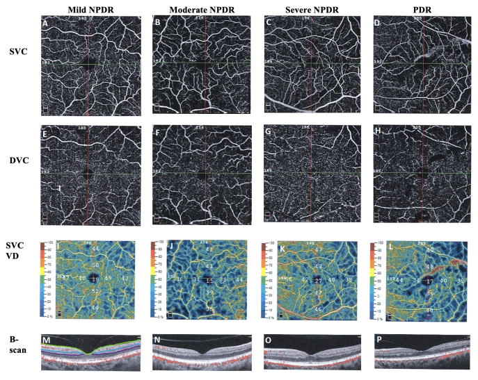
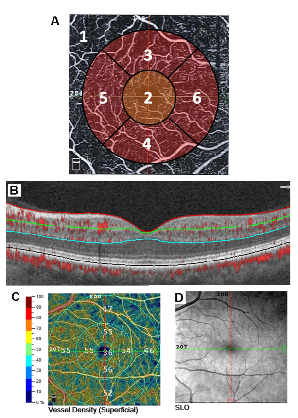
Methods: This retrospective cohort study analyzed the automatically quantified VDs of the superficial vascular complex (SVC) and deep vascular complex (DVC), including the whole, foveal, and parafoveal VDs, on quality OCT-A scans in patients diagnosed with DR. A multivariate linear regression and analysis of variance (ANOVA) analysis compared VDs to DR severity, visual acuity, and demographic factors. A linear mixed analysis determined the effects of VD by whether anti-VEGF therapy was given to patients with OCT-A scans at multiple time points.
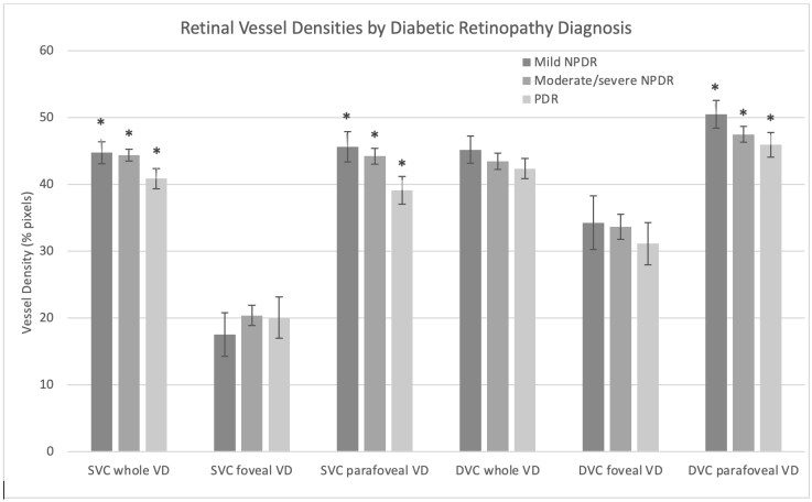
Results: There was a positive correlation of the VDs in both the SVC whole and parafoveal VD and DVC parafoveal VD with decreased DR severity and increased visual acuity (p≤0.001). The DVC whole VD was also positively correlated with increased visual acuity (p<0.001). There was no difference in the VDs associated with anti-VEGF treatment over time.
Conclusions: OCT-A VD shows promise for diagnosing and monitoring DR using DR severity and visual acuity. Anti-VEGF treatment had no significant effect (p=0.063) on vascular density in diabetic retinopathy.
期刊介绍:
Molecular Vision is a peer-reviewed journal dedicated to the dissemination of research results in molecular biology, cell biology, and the genetics of the visual system (ocular and cortical).
Molecular Vision publishes articles presenting original research that has not previously been published and comprehensive articles reviewing the current status of a particular field or topic. Submissions to Molecular Vision are subjected to rigorous peer review. Molecular Vision does NOT publish preprints.
For authors, Molecular Vision provides a rapid means of communicating important results. Access to Molecular Vision is free and unrestricted, allowing the widest possible audience for your article. Digital publishing allows you to use color images freely (and without fees). Additionally, you may publish animations, sounds, or other supplementary information that clarifies or supports your article. Each of the authors of an article may also list an electronic mail address (which will be updated upon request) to give interested readers easy access to authors.
 求助内容:
求助内容: 应助结果提醒方式:


