胃克罗恩病的一个不寻常的内镜发现。

IF 1 Q4 GASTROENTEROLOGY & HEPATOLOGY
Isabel Garrido, João Santos-Antunes, Guilherme Macedo
{"title":"胃克罗恩病的一个不寻常的内镜发现。","authors":"Isabel Garrido, João Santos-Antunes, Guilherme Macedo","doi":"10.1159/000520907","DOIUrl":null,"url":null,"abstract":"A 37-year-old woman with a past medical history of obesity (body mass index 50 kg/m2) and Crohn’s disease (Montreal classification – A2 L2+L4 B1) was referred to our institution for bariatric surgery after several unsuccessful weight loss attempts. The diagnosis of Crohn’s disease was established at age 19 (Fig. 1) and at that time she started therapy with azathioprine and a prednisolone taper. Due to poor control of the disease, the patient underwent several long courses of corticosteroid therapy. At this point, she began to gradually increase her weight. Over the past 4 years, the patient has been on infliximab at 5 mg/kg every 8 weeks, with evidence of clinical and endoscopic remission. A preoperative esophagogastroduodenoscopy was requested in order to assess the upper gastrointestinal tract for any abnormal findings as well as the presence of Helicobacter pylori (HP) infection. The endoscopy revealed retracted scar tissue in the gastric body (Fig. 2a) and antrum (Fig. 2b), with lesions with a bamboo-joint-like appearance and several pseudopolyps (Fig. 3). The esophagus and duodenum appeared normal. Histopathologic examination of biopsies from the stomach demonstrated mild foveolar hyperplasia and HP-negative chronic gastritis, with no signs of activity. No epithelioid granulomas, glandular atrophy, dysplasia, or signs of malignancy were identified. The patient underwent a sleeve gastrectomy without complications. The surgical specimen did not reveal any specific sign for inactive Crohn’s disease. Indeed, histopathological evaluation of the surgical specimen showed chronic gastritis and hyperplasia of the foveolar epithelium cells, without active inflammation, granulomas, dysplasia, or signs of malignancy. Five months after the surgery, she has already lost 36 kg and Crohn’s disease remains in remission. Crohn’s disease is defined by chronic inflammation that may involve any site of the gastrointestinal tract.","PeriodicalId":51838,"journal":{"name":"GE Portuguese Journal of Gastroenterology","volume":null,"pages":null},"PeriodicalIF":1.0000,"publicationDate":"2023-03-01","publicationTypes":"Journal Article","fieldsOfStudy":null,"isOpenAccess":false,"openAccessPdf":"https://ftp.ncbi.nlm.nih.gov/pub/pmc/oa_pdf/7a/99/pjg-0030-0166.PMC10050835.pdf","citationCount":"1","resultStr":"{\"title\":\"An Unusual Endoscopic Finding of Gastric Crohn's Disease.\",\"authors\":\"Isabel Garrido, João Santos-Antunes, Guilherme Macedo\",\"doi\":\"10.1159/000520907\",\"DOIUrl\":null,\"url\":null,\"abstract\":\"A 37-year-old woman with a past medical history of obesity (body mass index 50 kg/m2) and Crohn’s disease (Montreal classification – A2 L2+L4 B1) was referred to our institution for bariatric surgery after several unsuccessful weight loss attempts. The diagnosis of Crohn’s disease was established at age 19 (Fig. 1) and at that time she started therapy with azathioprine and a prednisolone taper. Due to poor control of the disease, the patient underwent several long courses of corticosteroid therapy. At this point, she began to gradually increase her weight. Over the past 4 years, the patient has been on infliximab at 5 mg/kg every 8 weeks, with evidence of clinical and endoscopic remission. A preoperative esophagogastroduodenoscopy was requested in order to assess the upper gastrointestinal tract for any abnormal findings as well as the presence of Helicobacter pylori (HP) infection. The endoscopy revealed retracted scar tissue in the gastric body (Fig. 2a) and antrum (Fig. 2b), with lesions with a bamboo-joint-like appearance and several pseudopolyps (Fig. 3). The esophagus and duodenum appeared normal. Histopathologic examination of biopsies from the stomach demonstrated mild foveolar hyperplasia and HP-negative chronic gastritis, with no signs of activity. No epithelioid granulomas, glandular atrophy, dysplasia, or signs of malignancy were identified. The patient underwent a sleeve gastrectomy without complications. The surgical specimen did not reveal any specific sign for inactive Crohn’s disease. Indeed, histopathological evaluation of the surgical specimen showed chronic gastritis and hyperplasia of the foveolar epithelium cells, without active inflammation, granulomas, dysplasia, or signs of malignancy. Five months after the surgery, she has already lost 36 kg and Crohn’s disease remains in remission. Crohn’s disease is defined by chronic inflammation that may involve any site of the gastrointestinal tract.\",\"PeriodicalId\":51838,\"journal\":{\"name\":\"GE Portuguese Journal of Gastroenterology\",\"volume\":null,\"pages\":null},\"PeriodicalIF\":1.0000,\"publicationDate\":\"2023-03-01\",\"publicationTypes\":\"Journal Article\",\"fieldsOfStudy\":null,\"isOpenAccess\":false,\"openAccessPdf\":\"https://ftp.ncbi.nlm.nih.gov/pub/pmc/oa_pdf/7a/99/pjg-0030-0166.PMC10050835.pdf\",\"citationCount\":\"1\",\"resultStr\":null,\"platform\":\"Semanticscholar\",\"paperid\":null,\"PeriodicalName\":\"GE Portuguese Journal of Gastroenterology\",\"FirstCategoryId\":\"1085\",\"ListUrlMain\":\"https://doi.org/10.1159/000520907\",\"RegionNum\":0,\"RegionCategory\":null,\"ArticlePicture\":[],\"TitleCN\":null,\"AbstractTextCN\":null,\"PMCID\":null,\"EPubDate\":\"\",\"PubModel\":\"\",\"JCR\":\"Q4\",\"JCRName\":\"GASTROENTEROLOGY & HEPATOLOGY\",\"Score\":null,\"Total\":0}","platform":"Semanticscholar","paperid":null,"PeriodicalName":"GE Portuguese Journal of Gastroenterology","FirstCategoryId":"1085","ListUrlMain":"https://doi.org/10.1159/000520907","RegionNum":0,"RegionCategory":null,"ArticlePicture":[],"TitleCN":null,"AbstractTextCN":null,"PMCID":null,"EPubDate":"","PubModel":"","JCR":"Q4","JCRName":"GASTROENTEROLOGY & HEPATOLOGY","Score":null,"Total":0}
引用次数: 1

摘要

本文章由计算机程序翻译,如有差异,请以英文原文为准。
An Unusual Endoscopic Finding of Gastric Crohn's Disease.

An Unusual Endoscopic Finding of Gastric Crohn's Disease.

An Unusual Endoscopic Finding of Gastric Crohn's Disease.

An Unusual Endoscopic Finding of Gastric Crohn's Disease.
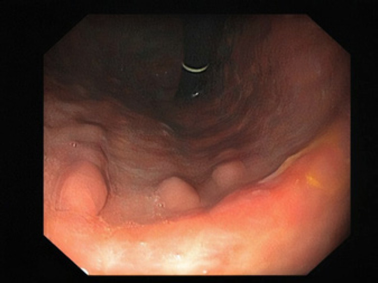
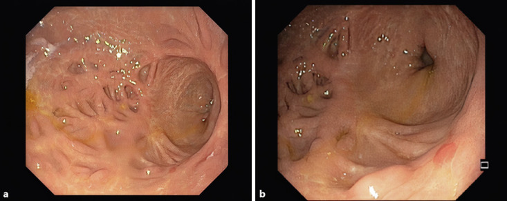
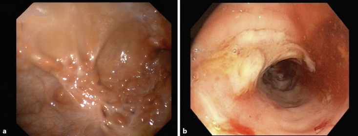
A 37-year-old woman with a past medical history of obesity (body mass index 50 kg/m2) and Crohn’s disease (Montreal classification – A2 L2+L4 B1) was referred to our institution for bariatric surgery after several unsuccessful weight loss attempts. The diagnosis of Crohn’s disease was established at age 19 (Fig. 1) and at that time she started therapy with azathioprine and a prednisolone taper. Due to poor control of the disease, the patient underwent several long courses of corticosteroid therapy. At this point, she began to gradually increase her weight. Over the past 4 years, the patient has been on infliximab at 5 mg/kg every 8 weeks, with evidence of clinical and endoscopic remission. A preoperative esophagogastroduodenoscopy was requested in order to assess the upper gastrointestinal tract for any abnormal findings as well as the presence of Helicobacter pylori (HP) infection. The endoscopy revealed retracted scar tissue in the gastric body (Fig. 2a) and antrum (Fig. 2b), with lesions with a bamboo-joint-like appearance and several pseudopolyps (Fig. 3). The esophagus and duodenum appeared normal. Histopathologic examination of biopsies from the stomach demonstrated mild foveolar hyperplasia and HP-negative chronic gastritis, with no signs of activity. No epithelioid granulomas, glandular atrophy, dysplasia, or signs of malignancy were identified. The patient underwent a sleeve gastrectomy without complications. The surgical specimen did not reveal any specific sign for inactive Crohn’s disease. Indeed, histopathological evaluation of the surgical specimen showed chronic gastritis and hyperplasia of the foveolar epithelium cells, without active inflammation, granulomas, dysplasia, or signs of malignancy. Five months after the surgery, she has already lost 36 kg and Crohn’s disease remains in remission. Crohn’s disease is defined by chronic inflammation that may involve any site of the gastrointestinal tract.
求助全文
通过发布文献求助,成功后即可免费获取论文全文。 去求助
来源期刊
GE Portuguese Journal of Gastroenterology
GE Portuguese Journal of Gastroenterology GASTROENTEROLOGY & HEPATOLOGY-
CiteScore
1.60
自引率
11.10%
发文量
62
审稿时长
21 weeks
期刊介绍: The ''GE Portuguese Journal of Gastroenterology'' (formerly Jornal Português de Gastrenterologia), founded in 1994, is the official publication of Sociedade Portuguesa de Gastrenterologia (Portuguese Society of Gastroenterology), Sociedade Portuguesa de Endoscopia Digestiva (Portuguese Society of Digestive Endoscopy) and Associação Portuguesa para o Estudo do Fígado (Portuguese Association for the Study of the Liver). The journal publishes clinical and basic research articles on Gastroenterology, Digestive Endoscopy, Hepatology and related topics. Review articles, clinical case studies, images, letters to the editor and other articles such as recommendations or papers on gastroenterology clinical practice are also considered. Only articles written in English are accepted.
×
引用
GB/T 7714-2015
复制
MLA
复制
APA
复制
导出至
BibTeX EndNote RefMan NoteFirst NoteExpress
×
提示
您的信息不完整,为了账户安全,请先补充。
现在去补充
×
提示
您因"违规操作"
具体请查看互助需知
我知道了
×
提示
确定
请完成安全验证×
copy
已复制链接
快去分享给好友吧!
我知道了
右上角分享
点击右上角分享
0
联系我们:info@booksci.cn Book学术提供免费学术资源搜索服务,方便国内外学者检索中英文文献。致力于提供最便捷和优质的服务体验。 Copyright © 2023 布克学术 All rights reserved.
京ICP备2023020795号-1
ghs 京公网安备 11010802042870号
Book学术文献互助
Book学术文献互助群
群 号:481959085
Book学术官方微信