{"title":"问题信息","authors":"","doi":"10.1002/cpcb.93","DOIUrl":null,"url":null,"abstract":"<p><b>Cover</b>: In Hegazy et al. (http://doi.org/10.1002/cpcb.115), the image shows the use of PLA to compare protein-protein interactions in clinical tissue specimens in situ. The interaction between the desmosomal cadherin desmoglein 1 (Dsg1<sup>goat</sup>) and its cytoplasmic partner plakoglobin (Pg<sup>mouse</sup>) is analyzed in a paraffin-embedded biopsy from a patient with SAM syndrome and compared to control tissue. SAM syndrome is a skin disease associated with a loss of Dsg1 expression at the cell-cell adhesive interface (Cohen-Barak et al., 2020; Samuelov et al., 2013). (<b>A</b>) The PLA signal is clearly decreased in the patient section compared to control tissue (red, Duolink™ In Situ Detection Reagent Red, λ<sub>Ex</sub>: 594, λ<sub>Em</sub>: 624). (<b>B</b>) A traditional immunofluorescence assay performed on control tissue is used to demonstrate successful antibody binding to both proteins analyzed by PLA (Dsg1, red; Pg, green; DAPI, blue). (<b>C</b>) In tissue sections such as this, using DAPI (blue) as a measure of cell number is not reliable as not every cell will have a nucleus in the cross section; therefore, the PLA signal is quantified and plotted as the area of PLA signal per field. Data points in the graph indicate the area of PLA signal in a region of the tissue. Error bars indicate the standard deviation. ImageJ/Fiji was used for image analysis and data graphed using GraphPad Prism<sup>®</sup>. Images were acquired using an AxioVison Z1 system (Carl Zeiss) with an Apotome slide module, an AxioCam MRm digital camera, and a 40× ([0.5 NA] Plan-Neofluar) objective. Scale bar, 50 µm.\n\n <figure>\n <div><picture>\n <source></source></picture><p></p>\n </div>\n </figure></p>","PeriodicalId":40051,"journal":{"name":"Current Protocols in Cell Biology","volume":"89 1","pages":""},"PeriodicalIF":0.0000,"publicationDate":"2020-12-22","publicationTypes":"Journal Article","fieldsOfStudy":null,"isOpenAccess":false,"openAccessPdf":"https://sci-hub-pdf.com/10.1002/cpcb.93","citationCount":"0","resultStr":"{\"title\":\"Issue Information\",\"authors\":\"\",\"doi\":\"10.1002/cpcb.93\",\"DOIUrl\":null,\"url\":null,\"abstract\":\"<p><b>Cover</b>: In Hegazy et al. (http://doi.org/10.1002/cpcb.115), the image shows the use of PLA to compare protein-protein interactions in clinical tissue specimens in situ. The interaction between the desmosomal cadherin desmoglein 1 (Dsg1<sup>goat</sup>) and its cytoplasmic partner plakoglobin (Pg<sup>mouse</sup>) is analyzed in a paraffin-embedded biopsy from a patient with SAM syndrome and compared to control tissue. SAM syndrome is a skin disease associated with a loss of Dsg1 expression at the cell-cell adhesive interface (Cohen-Barak et al., 2020; Samuelov et al., 2013). (<b>A</b>) The PLA signal is clearly decreased in the patient section compared to control tissue (red, Duolink™ In Situ Detection Reagent Red, λ<sub>Ex</sub>: 594, λ<sub>Em</sub>: 624). (<b>B</b>) A traditional immunofluorescence assay performed on control tissue is used to demonstrate successful antibody binding to both proteins analyzed by PLA (Dsg1, red; Pg, green; DAPI, blue). (<b>C</b>) In tissue sections such as this, using DAPI (blue) as a measure of cell number is not reliable as not every cell will have a nucleus in the cross section; therefore, the PLA signal is quantified and plotted as the area of PLA signal per field. Data points in the graph indicate the area of PLA signal in a region of the tissue. Error bars indicate the standard deviation. ImageJ/Fiji was used for image analysis and data graphed using GraphPad Prism<sup>®</sup>. Images were acquired using an AxioVison Z1 system (Carl Zeiss) with an Apotome slide module, an AxioCam MRm digital camera, and a 40× ([0.5 NA] Plan-Neofluar) objective. Scale bar, 50 µm.\\n\\n <figure>\\n <div><picture>\\n <source></source></picture><p></p>\\n </div>\\n </figure></p>\",\"PeriodicalId\":40051,\"journal\":{\"name\":\"Current Protocols in Cell Biology\",\"volume\":\"89 1\",\"pages\":\"\"},\"PeriodicalIF\":0.0000,\"publicationDate\":\"2020-12-22\",\"publicationTypes\":\"Journal Article\",\"fieldsOfStudy\":null,\"isOpenAccess\":false,\"openAccessPdf\":\"https://sci-hub-pdf.com/10.1002/cpcb.93\",\"citationCount\":\"0\",\"resultStr\":null,\"platform\":\"Semanticscholar\",\"paperid\":null,\"PeriodicalName\":\"Current Protocols in Cell Biology\",\"FirstCategoryId\":\"1085\",\"ListUrlMain\":\"https://onlinelibrary.wiley.com/doi/10.1002/cpcb.93\",\"RegionNum\":0,\"RegionCategory\":null,\"ArticlePicture\":[],\"TitleCN\":null,\"AbstractTextCN\":null,\"PMCID\":null,\"EPubDate\":\"\",\"PubModel\":\"\",\"JCR\":\"Q3\",\"JCRName\":\"Biochemistry, Genetics and Molecular Biology\",\"Score\":null,\"Total\":0}","platform":"Semanticscholar","paperid":null,"PeriodicalName":"Current Protocols in Cell Biology","FirstCategoryId":"1085","ListUrlMain":"https://onlinelibrary.wiley.com/doi/10.1002/cpcb.93","RegionNum":0,"RegionCategory":null,"ArticlePicture":[],"TitleCN":null,"AbstractTextCN":null,"PMCID":null,"EPubDate":"","PubModel":"","JCR":"Q3","JCRName":"Biochemistry, Genetics and Molecular Biology","Score":null,"Total":0}
引用次数: 0
摘要
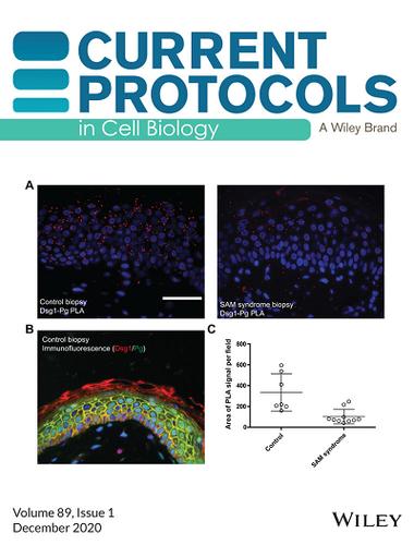
封面:在Hegazy等人(http://doi.org/10.1002/cpcb.115)中,该图像显示了使用聚乳酸来原位比较临床组织标本中蛋白质-蛋白质相互作用。在SAM综合征患者的石蜡包埋活检中分析了桥粒钙粘蛋白桥粒蛋白1 (Dsg1goat)与其细胞质伙伴血小板红蛋白(Pgmouse)之间的相互作用,并与对照组织进行了比较。SAM综合征是一种与细胞-细胞粘附界面Dsg1表达缺失相关的皮肤病(Cohen-Barak et al., 2020;Samuelov et al., 2013)。(A)与对照组织相比,患者切片中的PLA信号明显降低(红色,Duolink™原位检测试剂红色,λEx: 594, λEm: 624)。(B)在对照组织上进行的传统免疫荧光试验证明抗体与PLA分析的两种蛋白质成功结合(Dsg1,红色;Pg,绿色;DAPI,蓝色)。(C)在这样的组织切片中,使用DAPI(蓝色)作为细胞数量的测量是不可靠的,因为不是每个细胞在横截面上都有细胞核;因此,PLA信号被量化并绘制为每个场的PLA信号面积。图中的数据点表示PLA信号在组织区域中的面积。误差条表示标准差。使用ImageJ/Fiji进行图像分析,并使用GraphPad Prism®绘制数据。使用带有apooome载玻片模块的AxioVison Z1系统(卡尔蔡司)、AxioCam MRm数码相机和40× ([0.5 NA] Plan-Neofluar)物镜获得图像。比例尺,50µm。
Cover: In Hegazy et al. (http://doi.org/10.1002/cpcb.115), the image shows the use of PLA to compare protein-protein interactions in clinical tissue specimens in situ. The interaction between the desmosomal cadherin desmoglein 1 (Dsg1goat) and its cytoplasmic partner plakoglobin (Pgmouse) is analyzed in a paraffin-embedded biopsy from a patient with SAM syndrome and compared to control tissue. SAM syndrome is a skin disease associated with a loss of Dsg1 expression at the cell-cell adhesive interface (Cohen-Barak et al., 2020; Samuelov et al., 2013). (A) The PLA signal is clearly decreased in the patient section compared to control tissue (red, Duolink™ In Situ Detection Reagent Red, λEx: 594, λEm: 624). (B) A traditional immunofluorescence assay performed on control tissue is used to demonstrate successful antibody binding to both proteins analyzed by PLA (Dsg1, red; Pg, green; DAPI, blue). (C) In tissue sections such as this, using DAPI (blue) as a measure of cell number is not reliable as not every cell will have a nucleus in the cross section; therefore, the PLA signal is quantified and plotted as the area of PLA signal per field. Data points in the graph indicate the area of PLA signal in a region of the tissue. Error bars indicate the standard deviation. ImageJ/Fiji was used for image analysis and data graphed using GraphPad Prism®. Images were acquired using an AxioVison Z1 system (Carl Zeiss) with an Apotome slide module, an AxioCam MRm digital camera, and a 40× ([0.5 NA] Plan-Neofluar) objective. Scale bar, 50 µm.
期刊介绍:
Developed by leading scientists in the field, Current Protocols in Cell Biology is an essential reference for researchers who study the relationship between specific molecules and genes and their location, function and structure at the cellular level. Updated every three months in all formats, CPCB is constantly evolving to keep pace with the very latest discoveries and developments.

 求助内容:
求助内容: 应助结果提醒方式:


