{"title":"发布信息TOC","authors":"","doi":"10.1002/cpph.51","DOIUrl":null,"url":null,"abstract":"<p><b>Cover</b>: In Marchelletta et al. (https://doi.org/10.1002/cpph.54), the image show human colonic organoids retain in vivo morphology and cellular composition. Representative images of hematoxylin and eosin (H&E) staining of sections from paraffin-embedded organoids reveal a closed unit of differentiated columnar cells that resemble cells along the crypt axis in human colonic histologic sections from explant tissue. (<b>A</b>, <b>B</b>) Nuclei (dark purple) rest on the basal side in both explant tissue and organoids. (<b>C-F</b>) Organoids retain the cellular composition, frequency, and spatial orientation of tissue-resident colonic epithelial cells, as revealed by immunohistochemistry staining (arrows and brown staining) for the goblet-cell marker mucin 2 (MUC2) (<b>C</b>, <b>D</b>) and the enteroendocrine marker chromogranin A (<b>E</b>, <b>F</b>). \n\n <figure>\n <div><picture>\n <source></source></picture><p></p>\n </div>\n </figure></p>","PeriodicalId":10871,"journal":{"name":"Current Protocols in Pharmacology","volume":"85 1","pages":""},"PeriodicalIF":0.0000,"publicationDate":"2019-06-18","publicationTypes":"Journal Article","fieldsOfStudy":null,"isOpenAccess":false,"openAccessPdf":"https://sci-hub-pdf.com/10.1002/cpph.51","citationCount":"0","resultStr":"{\"title\":\"Issue Information TOC\",\"authors\":\"\",\"doi\":\"10.1002/cpph.51\",\"DOIUrl\":null,\"url\":null,\"abstract\":\"<p><b>Cover</b>: In Marchelletta et al. (https://doi.org/10.1002/cpph.54), the image show human colonic organoids retain in vivo morphology and cellular composition. Representative images of hematoxylin and eosin (H&E) staining of sections from paraffin-embedded organoids reveal a closed unit of differentiated columnar cells that resemble cells along the crypt axis in human colonic histologic sections from explant tissue. (<b>A</b>, <b>B</b>) Nuclei (dark purple) rest on the basal side in both explant tissue and organoids. (<b>C-F</b>) Organoids retain the cellular composition, frequency, and spatial orientation of tissue-resident colonic epithelial cells, as revealed by immunohistochemistry staining (arrows and brown staining) for the goblet-cell marker mucin 2 (MUC2) (<b>C</b>, <b>D</b>) and the enteroendocrine marker chromogranin A (<b>E</b>, <b>F</b>). \\n\\n <figure>\\n <div><picture>\\n <source></source></picture><p></p>\\n </div>\\n </figure></p>\",\"PeriodicalId\":10871,\"journal\":{\"name\":\"Current Protocols in Pharmacology\",\"volume\":\"85 1\",\"pages\":\"\"},\"PeriodicalIF\":0.0000,\"publicationDate\":\"2019-06-18\",\"publicationTypes\":\"Journal Article\",\"fieldsOfStudy\":null,\"isOpenAccess\":false,\"openAccessPdf\":\"https://sci-hub-pdf.com/10.1002/cpph.51\",\"citationCount\":\"0\",\"resultStr\":null,\"platform\":\"Semanticscholar\",\"paperid\":null,\"PeriodicalName\":\"Current Protocols in Pharmacology\",\"FirstCategoryId\":\"1085\",\"ListUrlMain\":\"https://onlinelibrary.wiley.com/doi/10.1002/cpph.51\",\"RegionNum\":0,\"RegionCategory\":null,\"ArticlePicture\":[],\"TitleCN\":null,\"AbstractTextCN\":null,\"PMCID\":null,\"EPubDate\":\"\",\"PubModel\":\"\",\"JCR\":\"Q2\",\"JCRName\":\"Pharmacology, Toxicology and Pharmaceutics\",\"Score\":null,\"Total\":0}","platform":"Semanticscholar","paperid":null,"PeriodicalName":"Current Protocols in Pharmacology","FirstCategoryId":"1085","ListUrlMain":"https://onlinelibrary.wiley.com/doi/10.1002/cpph.51","RegionNum":0,"RegionCategory":null,"ArticlePicture":[],"TitleCN":null,"AbstractTextCN":null,"PMCID":null,"EPubDate":"","PubModel":"","JCR":"Q2","JCRName":"Pharmacology, Toxicology and Pharmaceutics","Score":null,"Total":0}
引用次数: 0
摘要
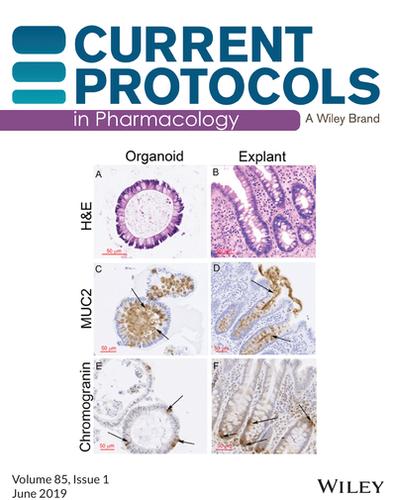
封面:在Marchelletta等人(https://doi.org/10.1002/cpph.54)中,图像显示人类结肠类器官保留了体内形态和细胞组成。石蜡包埋的类器官切片的代表性图像苏木精和伊红(H&E)染色显示一个封闭的分化柱状细胞单位,类似于来自外植体组织的人类结肠组织学切片中沿隐窝轴的细胞。(A, B)在外植体组织和类器官中,细胞核(深紫色)位于基侧。(C-F)免疫组织化学染色(箭头和棕色染色)显示,类器官保留了组织驻留的结肠上皮细胞的细胞组成、频率和空间取向,如杯状细胞标记mucin 2 (C, D)和肠内分泌标记chromogranin A (E, F)。
Cover: In Marchelletta et al. (https://doi.org/10.1002/cpph.54), the image show human colonic organoids retain in vivo morphology and cellular composition. Representative images of hematoxylin and eosin (H&E) staining of sections from paraffin-embedded organoids reveal a closed unit of differentiated columnar cells that resemble cells along the crypt axis in human colonic histologic sections from explant tissue. (A, B) Nuclei (dark purple) rest on the basal side in both explant tissue and organoids. (C-F) Organoids retain the cellular composition, frequency, and spatial orientation of tissue-resident colonic epithelial cells, as revealed by immunohistochemistry staining (arrows and brown staining) for the goblet-cell marker mucin 2 (MUC2) (C, D) and the enteroendocrine marker chromogranin A (E, F).

 求助内容:
求助内容: 应助结果提醒方式:


