{"title":"多组学分析显示,β-肌球蛋白重链表达的心肌细胞在早期心肌肥厚中糖酵解过程丰富","authors":"Hsiao-hui Yeh , Yao-Ming Chang , Yu-Wang Chang , Mei-Yeh Jade Lu , Yi-Hua Chen , Chia-Che Lee , Chien-Chang Chen","doi":"10.1016/j.jmccpl.2022.100011","DOIUrl":null,"url":null,"abstract":"<div><h3>Background</h3><p>Cardiac pressure overload induces cardiac hypertrophy and eventually leads to heart failure. One distinct feature of pathological cardiac hypertrophy is fetal-gene re-expression, but not every cardiomyocyte exhibits fetal gene re-expression in the diseased heart. Adult cardiomyocytes are terminally differentiated cells, so we do not know how the heterogeneity is determined and whether the differential fetal-gene reprogramming indicates a different degree of remodeling among cardiomyocytes. We hypothesized that fetal gene-expressed cardiomyocytes show more pathological features in the pressure-overloaded heart.</p></div><div><h3>Results</h3><p>We induced pressure overload in mice by transverse aortic constriction (TAC) and observed a cardiomyocyte population with expression of β-myosin heavy chain (βMHC, a fetal gene encoded by <em>Myh7</em>) after TAC for 3 days. On transcriptomic and proteomic analyses, βMHC-expressed cardiomyocytes of 3-day TAC hearts were enriched in genes in cardiomyopathy-associated pathways and glycolytic processes. Moreover, results of immunoblotting and enzyme activity assay suggested higher glycolytic activity in βMHC-expressed than non-expressed cardiomyocytes. When we inhibited the glycolytic flux by 2-deoxy-<span>d</span>-glucose, a widely used glycolysis inhibitor, the number of βMHC-expressed cardiomyocytes was reduced, and the level of TEA domain family member 1 (TEAD1), a transcriptional enhancer, was decreased. Also, our spatial transcriptomic results demonstrated that naïve and 3-day TAC hearts had fetal-gene–rich tissue domains that were enriched in pathways in extracellular matrix organization and tissue remodeling. As well, gene levels of glycolytic enzymes were higher in <em>Myh7</em>-positive than <em>Myh7</em>-negative domains.</p></div><div><h3>Conclusions</h3><p>Our data suggest that βMHC-expressed cardiomyocytes progress to pathological remodeling in the early stages of cardiac hypertrophy. In addition, the diverse glycolytic activity among cardiomyocytes might play a role in regulating gene expression via TEAD1 signaling.</p></div>","PeriodicalId":73835,"journal":{"name":"Journal of molecular and cellular cardiology plus","volume":null,"pages":null},"PeriodicalIF":0.0000,"publicationDate":"2022-09-01","publicationTypes":"Journal Article","fieldsOfStudy":null,"isOpenAccess":false,"openAccessPdf":"https://www.sciencedirect.com/science/article/pii/S2772976122000058/pdfft?md5=33ea72a7a407ecf57335d0fa20f5f476&pid=1-s2.0-S2772976122000058-main.pdf","citationCount":"0","resultStr":"{\"title\":\"Multiomic analyses reveal enriched glycolytic processes in β-myosin heavy chain-expressed cardiomyocytes in early cardiac hypertrophy\",\"authors\":\"Hsiao-hui Yeh , Yao-Ming Chang , Yu-Wang Chang , Mei-Yeh Jade Lu , Yi-Hua Chen , Chia-Che Lee , Chien-Chang Chen\",\"doi\":\"10.1016/j.jmccpl.2022.100011\",\"DOIUrl\":null,\"url\":null,\"abstract\":\"<div><h3>Background</h3><p>Cardiac pressure overload induces cardiac hypertrophy and eventually leads to heart failure. One distinct feature of pathological cardiac hypertrophy is fetal-gene re-expression, but not every cardiomyocyte exhibits fetal gene re-expression in the diseased heart. Adult cardiomyocytes are terminally differentiated cells, so we do not know how the heterogeneity is determined and whether the differential fetal-gene reprogramming indicates a different degree of remodeling among cardiomyocytes. We hypothesized that fetal gene-expressed cardiomyocytes show more pathological features in the pressure-overloaded heart.</p></div><div><h3>Results</h3><p>We induced pressure overload in mice by transverse aortic constriction (TAC) and observed a cardiomyocyte population with expression of β-myosin heavy chain (βMHC, a fetal gene encoded by <em>Myh7</em>) after TAC for 3 days. On transcriptomic and proteomic analyses, βMHC-expressed cardiomyocytes of 3-day TAC hearts were enriched in genes in cardiomyopathy-associated pathways and glycolytic processes. Moreover, results of immunoblotting and enzyme activity assay suggested higher glycolytic activity in βMHC-expressed than non-expressed cardiomyocytes. When we inhibited the glycolytic flux by 2-deoxy-<span>d</span>-glucose, a widely used glycolysis inhibitor, the number of βMHC-expressed cardiomyocytes was reduced, and the level of TEA domain family member 1 (TEAD1), a transcriptional enhancer, was decreased. Also, our spatial transcriptomic results demonstrated that naïve and 3-day TAC hearts had fetal-gene–rich tissue domains that were enriched in pathways in extracellular matrix organization and tissue remodeling. As well, gene levels of glycolytic enzymes were higher in <em>Myh7</em>-positive than <em>Myh7</em>-negative domains.</p></div><div><h3>Conclusions</h3><p>Our data suggest that βMHC-expressed cardiomyocytes progress to pathological remodeling in the early stages of cardiac hypertrophy. In addition, the diverse glycolytic activity among cardiomyocytes might play a role in regulating gene expression via TEAD1 signaling.</p></div>\",\"PeriodicalId\":73835,\"journal\":{\"name\":\"Journal of molecular and cellular cardiology plus\",\"volume\":null,\"pages\":null},\"PeriodicalIF\":0.0000,\"publicationDate\":\"2022-09-01\",\"publicationTypes\":\"Journal Article\",\"fieldsOfStudy\":null,\"isOpenAccess\":false,\"openAccessPdf\":\"https://www.sciencedirect.com/science/article/pii/S2772976122000058/pdfft?md5=33ea72a7a407ecf57335d0fa20f5f476&pid=1-s2.0-S2772976122000058-main.pdf\",\"citationCount\":\"0\",\"resultStr\":null,\"platform\":\"Semanticscholar\",\"paperid\":null,\"PeriodicalName\":\"Journal of molecular and cellular cardiology plus\",\"FirstCategoryId\":\"1085\",\"ListUrlMain\":\"https://www.sciencedirect.com/science/article/pii/S2772976122000058\",\"RegionNum\":0,\"RegionCategory\":null,\"ArticlePicture\":[],\"TitleCN\":null,\"AbstractTextCN\":null,\"PMCID\":null,\"EPubDate\":\"\",\"PubModel\":\"\",\"JCR\":\"\",\"JCRName\":\"\",\"Score\":null,\"Total\":0}","platform":"Semanticscholar","paperid":null,"PeriodicalName":"Journal of molecular and cellular cardiology plus","FirstCategoryId":"1085","ListUrlMain":"https://www.sciencedirect.com/science/article/pii/S2772976122000058","RegionNum":0,"RegionCategory":null,"ArticlePicture":[],"TitleCN":null,"AbstractTextCN":null,"PMCID":null,"EPubDate":"","PubModel":"","JCR":"","JCRName":"","Score":null,"Total":0}
Multiomic analyses reveal enriched glycolytic processes in β-myosin heavy chain-expressed cardiomyocytes in early cardiac hypertrophy
Background
Cardiac pressure overload induces cardiac hypertrophy and eventually leads to heart failure. One distinct feature of pathological cardiac hypertrophy is fetal-gene re-expression, but not every cardiomyocyte exhibits fetal gene re-expression in the diseased heart. Adult cardiomyocytes are terminally differentiated cells, so we do not know how the heterogeneity is determined and whether the differential fetal-gene reprogramming indicates a different degree of remodeling among cardiomyocytes. We hypothesized that fetal gene-expressed cardiomyocytes show more pathological features in the pressure-overloaded heart.
Results
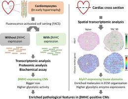
We induced pressure overload in mice by transverse aortic constriction (TAC) and observed a cardiomyocyte population with expression of β-myosin heavy chain (βMHC, a fetal gene encoded by Myh7) after TAC for 3 days. On transcriptomic and proteomic analyses, βMHC-expressed cardiomyocytes of 3-day TAC hearts were enriched in genes in cardiomyopathy-associated pathways and glycolytic processes. Moreover, results of immunoblotting and enzyme activity assay suggested higher glycolytic activity in βMHC-expressed than non-expressed cardiomyocytes. When we inhibited the glycolytic flux by 2-deoxy-d-glucose, a widely used glycolysis inhibitor, the number of βMHC-expressed cardiomyocytes was reduced, and the level of TEA domain family member 1 (TEAD1), a transcriptional enhancer, was decreased. Also, our spatial transcriptomic results demonstrated that naïve and 3-day TAC hearts had fetal-gene–rich tissue domains that were enriched in pathways in extracellular matrix organization and tissue remodeling. As well, gene levels of glycolytic enzymes were higher in Myh7-positive than Myh7-negative domains.
Conclusions
Our data suggest that βMHC-expressed cardiomyocytes progress to pathological remodeling in the early stages of cardiac hypertrophy. In addition, the diverse glycolytic activity among cardiomyocytes might play a role in regulating gene expression via TEAD1 signaling.

 求助内容:
求助内容: 应助结果提醒方式:


