Xiaojuan Li, Frank W Roemer, Flavia Cicuttini, Jamie W MacKay, Tom Turmezei, Thomas M Link
{"title":"早期膝关节 OA 的定义--现阶段我们知道些什么?影像学视角。","authors":"Xiaojuan Li, Frank W Roemer, Flavia Cicuttini, Jamie W MacKay, Tom Turmezei, Thomas M Link","doi":"10.1177/1759720X231158204","DOIUrl":null,"url":null,"abstract":"<p><p>While criteria for early-stage knee osteoarthritis (OA) in a primary care setting have been proposed, the role of imaging has been limited to radiography using the standard Kellgren-Lawrence classification. Standardized imaging and interpretation are critical with radiographs, yet studies have also shown that even early stages of radiographic OA already demonstrate advanced damage to knee joint tissues such as cartilage, menisci, and bone marrow. Morphological magnetic resonance imaging (MRI) shows degenerative damage earlier than radiographs and definitions for OA using MRI have been published though no accepted definition of early OA based on MRI is currently available. The clinical significance of structural abnormalities has also not been well defined, and the differentiation between normal aging and structural OA development remains a challenge. Compositional MRI of cartilage provides information on biochemical, degenerative changes within the cartilage matrix before cartilage defects occur and when cartilage damage is potentially reversible. Studies have shown that cartilage composition can predict cartilage loss and radiographic OA. However, while this technology is most promising for characterizing early OA it has currently limited clinical application. Better standardization of compositional MRI is required, which is currently work in progress. Finally, there has been renewed interest in computed tomography (CT) for assessing early knee OA as new techniques such as weight bearing and spectral CT are available, which may provide information on joint loading, cartilage, and bone and potentially have a role in better characterizing early OA. In conclusion, while imaging may have a limited role in diagnosing early OA in a primary care setting, there are advanced imaging technologies available, which detect early degeneration and may thus significantly alter management as new therapeutic modalities evolve.</p>","PeriodicalId":23056,"journal":{"name":"Therapeutic Advances in Musculoskeletal Disease","volume":"15 ","pages":"1759720X231158204"},"PeriodicalIF":4.1000,"publicationDate":"2023-03-14","publicationTypes":"Journal Article","fieldsOfStudy":null,"isOpenAccess":false,"openAccessPdf":"https://ftp.ncbi.nlm.nih.gov/pub/pmc/oa_pdf/f9/22/10.1177_1759720X231158204.PMC10017942.pdf","citationCount":"0","resultStr":"{\"title\":\"Early knee OA definition-what do we know at this stage? An imaging perspective.\",\"authors\":\"Xiaojuan Li, Frank W Roemer, Flavia Cicuttini, Jamie W MacKay, Tom Turmezei, Thomas M Link\",\"doi\":\"10.1177/1759720X231158204\",\"DOIUrl\":null,\"url\":null,\"abstract\":\"<p><p>While criteria for early-stage knee osteoarthritis (OA) in a primary care setting have been proposed, the role of imaging has been limited to radiography using the standard Kellgren-Lawrence classification. Standardized imaging and interpretation are critical with radiographs, yet studies have also shown that even early stages of radiographic OA already demonstrate advanced damage to knee joint tissues such as cartilage, menisci, and bone marrow. Morphological magnetic resonance imaging (MRI) shows degenerative damage earlier than radiographs and definitions for OA using MRI have been published though no accepted definition of early OA based on MRI is currently available. The clinical significance of structural abnormalities has also not been well defined, and the differentiation between normal aging and structural OA development remains a challenge. Compositional MRI of cartilage provides information on biochemical, degenerative changes within the cartilage matrix before cartilage defects occur and when cartilage damage is potentially reversible. Studies have shown that cartilage composition can predict cartilage loss and radiographic OA. However, while this technology is most promising for characterizing early OA it has currently limited clinical application. Better standardization of compositional MRI is required, which is currently work in progress. Finally, there has been renewed interest in computed tomography (CT) for assessing early knee OA as new techniques such as weight bearing and spectral CT are available, which may provide information on joint loading, cartilage, and bone and potentially have a role in better characterizing early OA. In conclusion, while imaging may have a limited role in diagnosing early OA in a primary care setting, there are advanced imaging technologies available, which detect early degeneration and may thus significantly alter management as new therapeutic modalities evolve.</p>\",\"PeriodicalId\":23056,\"journal\":{\"name\":\"Therapeutic Advances in Musculoskeletal Disease\",\"volume\":\"15 \",\"pages\":\"1759720X231158204\"},\"PeriodicalIF\":4.1000,\"publicationDate\":\"2023-03-14\",\"publicationTypes\":\"Journal Article\",\"fieldsOfStudy\":null,\"isOpenAccess\":false,\"openAccessPdf\":\"https://ftp.ncbi.nlm.nih.gov/pub/pmc/oa_pdf/f9/22/10.1177_1759720X231158204.PMC10017942.pdf\",\"citationCount\":\"0\",\"resultStr\":null,\"platform\":\"Semanticscholar\",\"paperid\":null,\"PeriodicalName\":\"Therapeutic Advances in Musculoskeletal Disease\",\"FirstCategoryId\":\"3\",\"ListUrlMain\":\"https://doi.org/10.1177/1759720X231158204\",\"RegionNum\":2,\"RegionCategory\":\"医学\",\"ArticlePicture\":[],\"TitleCN\":null,\"AbstractTextCN\":null,\"PMCID\":null,\"EPubDate\":\"2023/1/1 0:00:00\",\"PubModel\":\"eCollection\",\"JCR\":\"Q2\",\"JCRName\":\"RHEUMATOLOGY\",\"Score\":null,\"Total\":0}","platform":"Semanticscholar","paperid":null,"PeriodicalName":"Therapeutic Advances in Musculoskeletal Disease","FirstCategoryId":"3","ListUrlMain":"https://doi.org/10.1177/1759720X231158204","RegionNum":2,"RegionCategory":"医学","ArticlePicture":[],"TitleCN":null,"AbstractTextCN":null,"PMCID":null,"EPubDate":"2023/1/1 0:00:00","PubModel":"eCollection","JCR":"Q2","JCRName":"RHEUMATOLOGY","Score":null,"Total":0}
引用次数: 0
摘要
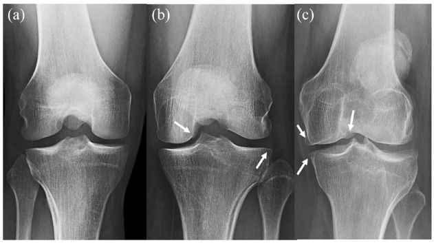
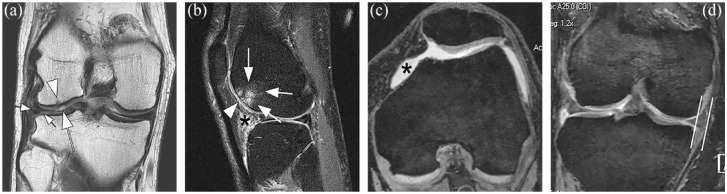
虽然已经提出了初级保健中早期膝关节骨性关节炎(OA)的标准,但影像学的作用仅限于使用标准的凯尔格伦-劳伦斯分类法进行放射摄影。标准化成像和判读对射线照相至关重要,但研究也表明,即使是早期的射线照相 OA 也已显示出膝关节组织(如软骨、半月板和骨髓)的晚期损伤。形态学核磁共振成像(MRI)比X光片更早显示出退行性损伤,尽管目前还没有基于核磁共振成像的早期 OA 的公认定义,但使用核磁共振成像对 OA 进行定义的研究已经发表。结构异常的临床意义也没有得到很好的界定,如何区分正常衰老和结构性 OA 的发展仍是一个挑战。软骨成分磁共振成像可在软骨缺损发生前和软骨损伤可能可逆时提供软骨基质内生化退行性变化的信息。研究表明,软骨成分可以预测软骨损失和放射学上的 OA。然而,虽然这项技术最有希望用于描述早期 OA,但目前的临床应用还很有限。需要对成分磁共振成像进行更好的标准化,目前这项工作正在进行中。最后,随着负重 CT 和频谱 CT 等新技术的出现,人们对用于评估早期膝关节 OA 的计算机断层扫描(CT)重新产生了兴趣。总之,虽然影像学在初级保健中诊断早期 OA 的作用有限,但目前已有先进的影像学技术可以检测早期退化,因此随着新治疗模式的发展,可能会大大改变治疗方法。
Early knee OA definition-what do we know at this stage? An imaging perspective.
While criteria for early-stage knee osteoarthritis (OA) in a primary care setting have been proposed, the role of imaging has been limited to radiography using the standard Kellgren-Lawrence classification. Standardized imaging and interpretation are critical with radiographs, yet studies have also shown that even early stages of radiographic OA already demonstrate advanced damage to knee joint tissues such as cartilage, menisci, and bone marrow. Morphological magnetic resonance imaging (MRI) shows degenerative damage earlier than radiographs and definitions for OA using MRI have been published though no accepted definition of early OA based on MRI is currently available. The clinical significance of structural abnormalities has also not been well defined, and the differentiation between normal aging and structural OA development remains a challenge. Compositional MRI of cartilage provides information on biochemical, degenerative changes within the cartilage matrix before cartilage defects occur and when cartilage damage is potentially reversible. Studies have shown that cartilage composition can predict cartilage loss and radiographic OA. However, while this technology is most promising for characterizing early OA it has currently limited clinical application. Better standardization of compositional MRI is required, which is currently work in progress. Finally, there has been renewed interest in computed tomography (CT) for assessing early knee OA as new techniques such as weight bearing and spectral CT are available, which may provide information on joint loading, cartilage, and bone and potentially have a role in better characterizing early OA. In conclusion, while imaging may have a limited role in diagnosing early OA in a primary care setting, there are advanced imaging technologies available, which detect early degeneration and may thus significantly alter management as new therapeutic modalities evolve.
期刊介绍:
Therapeutic Advances in Musculoskeletal Disease delivers the highest quality peer-reviewed articles, reviews, and scholarly comment on pioneering efforts and innovative studies across all areas of musculoskeletal disease.

 求助内容:
求助内容: 应助结果提醒方式:


