利用空气扫描共聚焦显微镜观察植物根部木质部导管中出现的次生增厚中的纤维素合酶
IF 6.2
Q1 Immunology and Microbiology
引用次数: 0
摘要
到目前为止,已经在植物表皮上观察到纤维素合酶颗粒的运动,这些颗粒适合共聚焦成像,产生可观的信号和分辨率来观察小的质膜局部颗粒。这里提出了一种方法,使用空气扫描共聚焦显微镜,允许在完整根的发育原木质部导管的深处获得类似的信息。本文章由计算机程序翻译,如有差异,请以英文原文为准。
Observing cellulose synthases at emerging secondary thickenings in developing xylem vessels of the plant root using airyscan confocal microscopy
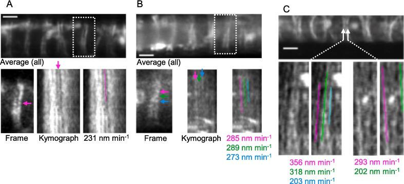
Movement of cellulose synthase particles have so far been observed on the plant epidermis that are amenable to confocal imaging, yielding appreciable signal and resolution to observe small plasma membrane-localised particles. Presented here is a method, using airyscan confocal microscopy, that permits similar information to be obtained at depth within the developing protoxylem vessels of intact roots.
求助全文
通过发布文献求助,成功后即可免费获取论文全文。
去求助
来源期刊

Cell Surface
Immunology and Microbiology-Applied Microbiology and Biotechnology
CiteScore
6.10
自引率
0.00%
发文量
18
审稿时长
49 days
 求助内容:
求助内容: 应助结果提醒方式:


