{"title":"大麻使用障碍的皮层形态:经颅直流电刺激治疗的意义。","authors":"Ghazaleh Soleimani, Farzad Towhidkhah, Mehrdad Saviz, Hamed Ekhtiari","doi":"10.32598/bcn.2021.3400.1","DOIUrl":null,"url":null,"abstract":"<p><strong>Introduction: </strong>Transcranial direct current stimulation (tDCS) has been studied as an adjunctive treatment option for substance use disorders (SUDs). Alterations in brain structure following SUD may change tDCS-induced electric field (EF) and subsequent responses; however, group-level differences between healthy controls (HC) and participants with SUDs in terms of EF and its association with cortical architecture have not yet been modeled quantitatively. This study provides a methodology for group-level analysis of computational head models to investigate the influence of cortical morphology metrics on EFs.</p><p><strong>Methods: </strong>Whole-brain surface-based morphology was conducted, and cortical thickness, volume, and surface area were compared between participants with cannabis use disorders (CUD) (n=20) and age-matched HC (n=22). Meanwhile, EFs were simulated for bilateral tDCS over the dorsolateral prefrontal cortex. The effects of structural alterations on EF distribution were investigated based on individualized computational head models.</p><p><strong>Results: </strong>Regarding EF, no significant difference was found within the prefrontal cortex; however, EFs were significantly different in left-postcentral and right-superior temporal gyrus (P<0.05) with higher levels of variance in CUD compared to HC [F<sub>(39, 43)</sub>=5.31, P<0.0001, C=0.95]. Significant differences were observed in cortical area (caudal anterior cingulate and rostral middle frontal), thickness (lateral orbitofrontal), and volume (paracentral and fusiform) between the two groups.</p><p><strong>Conclusion: </strong>Brain morphology and tDCS-induced EFs may be changed following CUD; however, differences between CUD and HCs in EFs do not always overlap with brain areas that show structural alterations. To sufficiently modulate stimulation targets, whether individuals with CUD need different stimulation doses based on tDCS target location should be checked.</p>","PeriodicalId":9311,"journal":{"name":"British Heart Journal","volume":"102 1","pages":"647-662"},"PeriodicalIF":0.0000,"publicationDate":"2023-09-01","publicationTypes":"Journal Article","fieldsOfStudy":null,"isOpenAccess":false,"openAccessPdf":"https://www.ncbi.nlm.nih.gov/pmc/articles/PMC11016884/pdf/","citationCount":"0","resultStr":"{\"title\":\"Cortical Morphology in Cannabis Use Disorder: Implications for Transcranial Direct Current Stimulation Treatment.\",\"authors\":\"Ghazaleh Soleimani, Farzad Towhidkhah, Mehrdad Saviz, Hamed Ekhtiari\",\"doi\":\"10.32598/bcn.2021.3400.1\",\"DOIUrl\":null,\"url\":null,\"abstract\":\"<p><strong>Introduction: </strong>Transcranial direct current stimulation (tDCS) has been studied as an adjunctive treatment option for substance use disorders (SUDs). Alterations in brain structure following SUD may change tDCS-induced electric field (EF) and subsequent responses; however, group-level differences between healthy controls (HC) and participants with SUDs in terms of EF and its association with cortical architecture have not yet been modeled quantitatively. This study provides a methodology for group-level analysis of computational head models to investigate the influence of cortical morphology metrics on EFs.</p><p><strong>Methods: </strong>Whole-brain surface-based morphology was conducted, and cortical thickness, volume, and surface area were compared between participants with cannabis use disorders (CUD) (n=20) and age-matched HC (n=22). Meanwhile, EFs were simulated for bilateral tDCS over the dorsolateral prefrontal cortex. The effects of structural alterations on EF distribution were investigated based on individualized computational head models.</p><p><strong>Results: </strong>Regarding EF, no significant difference was found within the prefrontal cortex; however, EFs were significantly different in left-postcentral and right-superior temporal gyrus (P<0.05) with higher levels of variance in CUD compared to HC [F<sub>(39, 43)</sub>=5.31, P<0.0001, C=0.95]. Significant differences were observed in cortical area (caudal anterior cingulate and rostral middle frontal), thickness (lateral orbitofrontal), and volume (paracentral and fusiform) between the two groups.</p><p><strong>Conclusion: </strong>Brain morphology and tDCS-induced EFs may be changed following CUD; however, differences between CUD and HCs in EFs do not always overlap with brain areas that show structural alterations. To sufficiently modulate stimulation targets, whether individuals with CUD need different stimulation doses based on tDCS target location should be checked.</p>\",\"PeriodicalId\":9311,\"journal\":{\"name\":\"British Heart Journal\",\"volume\":\"102 1\",\"pages\":\"647-662\"},\"PeriodicalIF\":0.0000,\"publicationDate\":\"2023-09-01\",\"publicationTypes\":\"Journal Article\",\"fieldsOfStudy\":null,\"isOpenAccess\":false,\"openAccessPdf\":\"https://www.ncbi.nlm.nih.gov/pmc/articles/PMC11016884/pdf/\",\"citationCount\":\"0\",\"resultStr\":null,\"platform\":\"Semanticscholar\",\"paperid\":null,\"PeriodicalName\":\"British Heart Journal\",\"FirstCategoryId\":\"1085\",\"ListUrlMain\":\"https://doi.org/10.32598/bcn.2021.3400.1\",\"RegionNum\":0,\"RegionCategory\":null,\"ArticlePicture\":[],\"TitleCN\":null,\"AbstractTextCN\":null,\"PMCID\":null,\"EPubDate\":\"\",\"PubModel\":\"\",\"JCR\":\"\",\"JCRName\":\"\",\"Score\":null,\"Total\":0}","platform":"Semanticscholar","paperid":null,"PeriodicalName":"British Heart Journal","FirstCategoryId":"1085","ListUrlMain":"https://doi.org/10.32598/bcn.2021.3400.1","RegionNum":0,"RegionCategory":null,"ArticlePicture":[],"TitleCN":null,"AbstractTextCN":null,"PMCID":null,"EPubDate":"","PubModel":"","JCR":"","JCRName":"","Score":null,"Total":0}
Cortical Morphology in Cannabis Use Disorder: Implications for Transcranial Direct Current Stimulation Treatment.
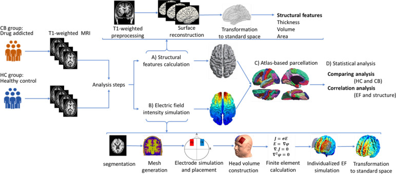
Introduction: Transcranial direct current stimulation (tDCS) has been studied as an adjunctive treatment option for substance use disorders (SUDs). Alterations in brain structure following SUD may change tDCS-induced electric field (EF) and subsequent responses; however, group-level differences between healthy controls (HC) and participants with SUDs in terms of EF and its association with cortical architecture have not yet been modeled quantitatively. This study provides a methodology for group-level analysis of computational head models to investigate the influence of cortical morphology metrics on EFs.
Methods: Whole-brain surface-based morphology was conducted, and cortical thickness, volume, and surface area were compared between participants with cannabis use disorders (CUD) (n=20) and age-matched HC (n=22). Meanwhile, EFs were simulated for bilateral tDCS over the dorsolateral prefrontal cortex. The effects of structural alterations on EF distribution were investigated based on individualized computational head models.
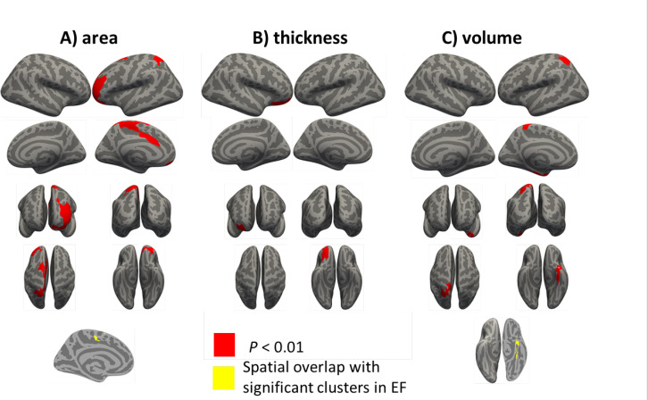
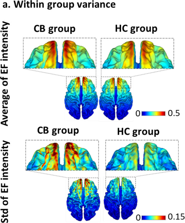
Results: Regarding EF, no significant difference was found within the prefrontal cortex; however, EFs were significantly different in left-postcentral and right-superior temporal gyrus (P<0.05) with higher levels of variance in CUD compared to HC [F(39, 43)=5.31, P<0.0001, C=0.95]. Significant differences were observed in cortical area (caudal anterior cingulate and rostral middle frontal), thickness (lateral orbitofrontal), and volume (paracentral and fusiform) between the two groups.
Conclusion: Brain morphology and tDCS-induced EFs may be changed following CUD; however, differences between CUD and HCs in EFs do not always overlap with brain areas that show structural alterations. To sufficiently modulate stimulation targets, whether individuals with CUD need different stimulation doses based on tDCS target location should be checked.

 求助内容:
求助内容: 应助结果提醒方式:


