S. Brugaletta, G. Giacchi, L. Ortega-Paz, H. M. Garcia-Garcia, M. Sabaté
{"title":"稳定的冠状动脉疾病。它真的稳定吗?冠状动脉病变的灰度和VH-IVUS形态学解释","authors":"S. Brugaletta, G. Giacchi, L. Ortega-Paz, H. M. Garcia-Garcia, M. Sabaté","doi":"10.1002/cce2.24","DOIUrl":null,"url":null,"abstract":"Despite the wide knowledge on the pathological features of an unstable plaque, it is hard to differentiate in vivo a stable from an unstable plaque. The possibility to differentiate these plaques is mainly based on coronary angiography, which depicts artery as a planar silhouette of the contrast‐filled lumen without any information on the vessel wall. Conversely, intracoronary imaging techniques, such as IVUS and IVUS virtual histology, are the established gold standard for in vivo qualitatively (e.g. visually) identification of plaque morphology and coronary plaque tissue components, helping understanding of plaque stability degree. We herein describe these imaging technologies and its clinical and research applications.","PeriodicalId":100331,"journal":{"name":"Continuing Cardiology Education","volume":"2 2","pages":"66-76"},"PeriodicalIF":0.0000,"publicationDate":"2016-06-22","publicationTypes":"Journal Article","fieldsOfStudy":null,"isOpenAccess":false,"openAccessPdf":"https://sci-hub-pdf.com/10.1002/cce2.24","citationCount":"4","resultStr":"{\"title\":\"Stable coronary artery disease. Is it really stable? Lesion morphology interpretation by Grayscale and VH-IVUS in patients with coronary artery disease\",\"authors\":\"S. Brugaletta, G. Giacchi, L. Ortega-Paz, H. M. Garcia-Garcia, M. Sabaté\",\"doi\":\"10.1002/cce2.24\",\"DOIUrl\":null,\"url\":null,\"abstract\":\"Despite the wide knowledge on the pathological features of an unstable plaque, it is hard to differentiate in vivo a stable from an unstable plaque. The possibility to differentiate these plaques is mainly based on coronary angiography, which depicts artery as a planar silhouette of the contrast‐filled lumen without any information on the vessel wall. Conversely, intracoronary imaging techniques, such as IVUS and IVUS virtual histology, are the established gold standard for in vivo qualitatively (e.g. visually) identification of plaque morphology and coronary plaque tissue components, helping understanding of plaque stability degree. We herein describe these imaging technologies and its clinical and research applications.\",\"PeriodicalId\":100331,\"journal\":{\"name\":\"Continuing Cardiology Education\",\"volume\":\"2 2\",\"pages\":\"66-76\"},\"PeriodicalIF\":0.0000,\"publicationDate\":\"2016-06-22\",\"publicationTypes\":\"Journal Article\",\"fieldsOfStudy\":null,\"isOpenAccess\":false,\"openAccessPdf\":\"https://sci-hub-pdf.com/10.1002/cce2.24\",\"citationCount\":\"4\",\"resultStr\":null,\"platform\":\"Semanticscholar\",\"paperid\":null,\"PeriodicalName\":\"Continuing Cardiology Education\",\"FirstCategoryId\":\"1085\",\"ListUrlMain\":\"https://onlinelibrary.wiley.com/doi/10.1002/cce2.24\",\"RegionNum\":0,\"RegionCategory\":null,\"ArticlePicture\":[],\"TitleCN\":null,\"AbstractTextCN\":null,\"PMCID\":null,\"EPubDate\":\"\",\"PubModel\":\"\",\"JCR\":\"\",\"JCRName\":\"\",\"Score\":null,\"Total\":0}","platform":"Semanticscholar","paperid":null,"PeriodicalName":"Continuing Cardiology Education","FirstCategoryId":"1085","ListUrlMain":"https://onlinelibrary.wiley.com/doi/10.1002/cce2.24","RegionNum":0,"RegionCategory":null,"ArticlePicture":[],"TitleCN":null,"AbstractTextCN":null,"PMCID":null,"EPubDate":"","PubModel":"","JCR":"","JCRName":"","Score":null,"Total":0}
Stable coronary artery disease. Is it really stable? Lesion morphology interpretation by Grayscale and VH-IVUS in patients with coronary artery disease
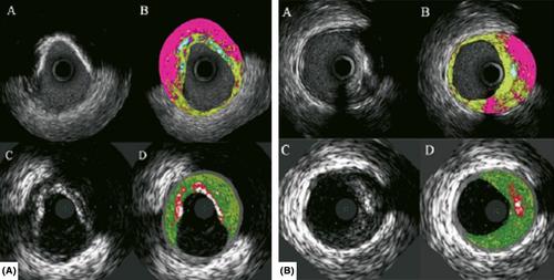
Despite the wide knowledge on the pathological features of an unstable plaque, it is hard to differentiate in vivo a stable from an unstable plaque. The possibility to differentiate these plaques is mainly based on coronary angiography, which depicts artery as a planar silhouette of the contrast‐filled lumen without any information on the vessel wall. Conversely, intracoronary imaging techniques, such as IVUS and IVUS virtual histology, are the established gold standard for in vivo qualitatively (e.g. visually) identification of plaque morphology and coronary plaque tissue components, helping understanding of plaque stability degree. We herein describe these imaging technologies and its clinical and research applications.

 求助内容:
求助内容: 应助结果提醒方式:


